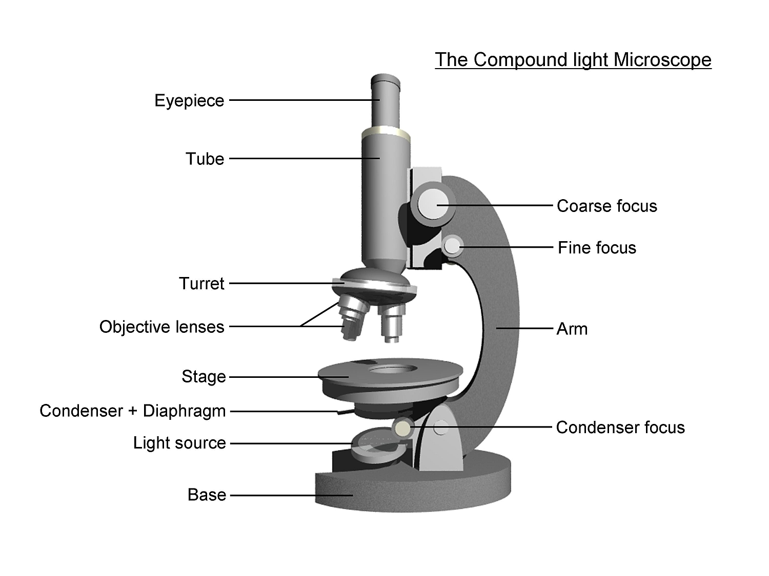
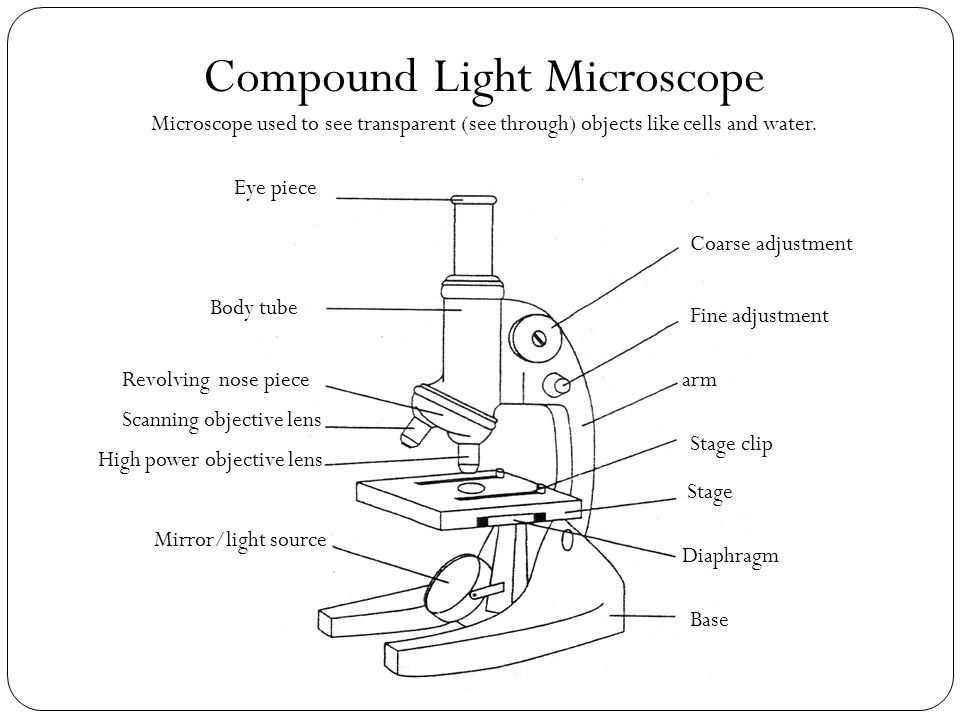
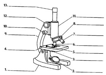
41 diagram of a compound light microscope
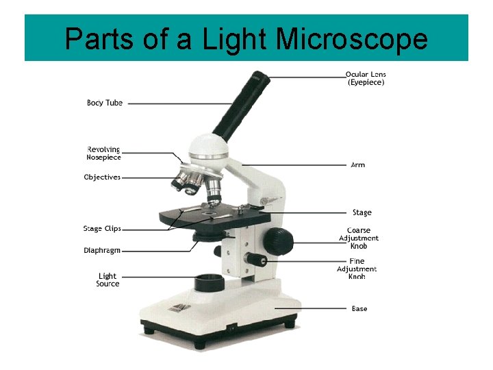
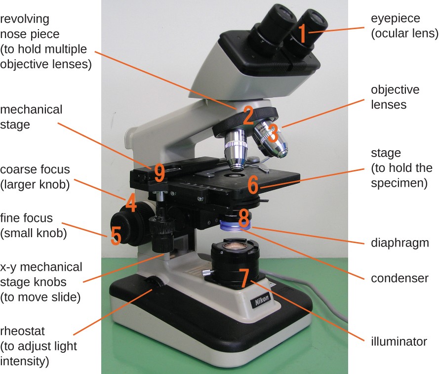
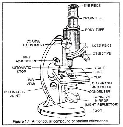
F. LIGHT SOURCE Projects light UPWARDS through the diaphragm, the SPECIMEN, and the LENSES H. DIAPHRAGM Regulates the amount of LIGHT on the specimen E. STAGE Supports the SLIDE being viewed K. ARM Used to SUPPORT the B. NOSEPIECE microscope when carried Holds the HIGH- and LOW- power objective LENSES; can be rotated to change MAGNIFICATION. Compound microscope is a type of optical microscope that is used for obtaining a high-resolution image. There are more than two lenses in a compound microscope. Learn about the working principle, parts and uses of a compound microscope along with a labeled diagram here.
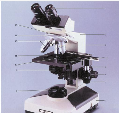
Parts of a microscope with functions and labeled diagram. Overview of Microscope and diagram. Structural parts of a Microscope and their functions. Optical ...

Diagram of a compound light microscope
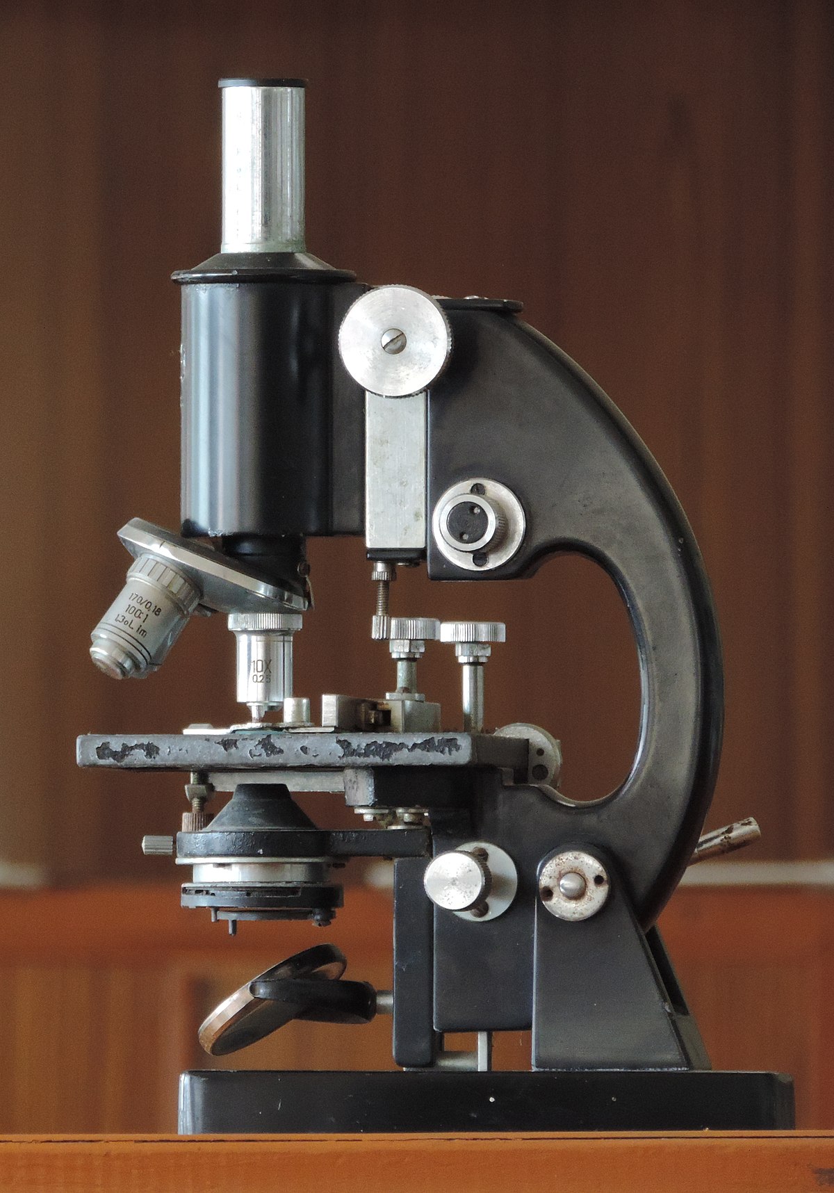
Simple light microscopes of the past could magnify an object to 266X as in the case of Leeuwenhoek's microscope. Modern compound light microscopes, under optimal conditions, can magnify an object from 1000X to 2000X (times) the specimens original diameter. Diagram Showing Light Traveling Through The Microscope Compound Microscope Diagram with Parts of The Support system. The Foot: It is the heavy metallic bottom part which supports all the other parts of the microscope. It may be oval, tripod or horseshoe shaped. Tube: It is the microscope arm's tubular, hollow component that is attached to the top half of the arm. It may be adjusted up and down ... Compound Microscope. A compound microscope uses a combination of lenses coupled with an artificial light source to magnify an object at various zoom levels to study the object. A compound microscope: Is used to view samples that are not visible to the naked eye; Uses two types of lenses - Objective and ocular lenses
Diagram of a compound light microscope. Microscope field of view 4000 urn The width (w) of this plant cell is closest to diagram below and on your knowledge of biology. The diagram shows cells as seen in the high-power (400> field of view of a compound light microscope. 0.5 mm What is the approximate diameter of cell A in micrometers? B) IOO C) 150 D) 250 I l. 15/05/2021 · Brightfield Microscope is also known as the Compound Light Microscope. It is an optical microscope that uses light rays to produce a dark image against a bright background. It is the standard microscope that is used in Biology, Cellular Biology, and Microbiological Laboratory studies. A compound microscope is the most common type of light (optical) microscopes. The term “compound” refers to the microscope having more than one lens. Basically, compound microscopes generate magnified images through an aligned pair of the objective lens and the ocular lens. The Optical Parts of Compound Microscope include: 1. Eyepiece lens or Ocular: At the top of the body tube, a lens is planted which is known as the eyepiece. On ...1 answer · Top answer: Hint: Magnification depends on focal length of objective and the eye lens. The least distance of distinct vision is 25 cm. The separation between the objective ...
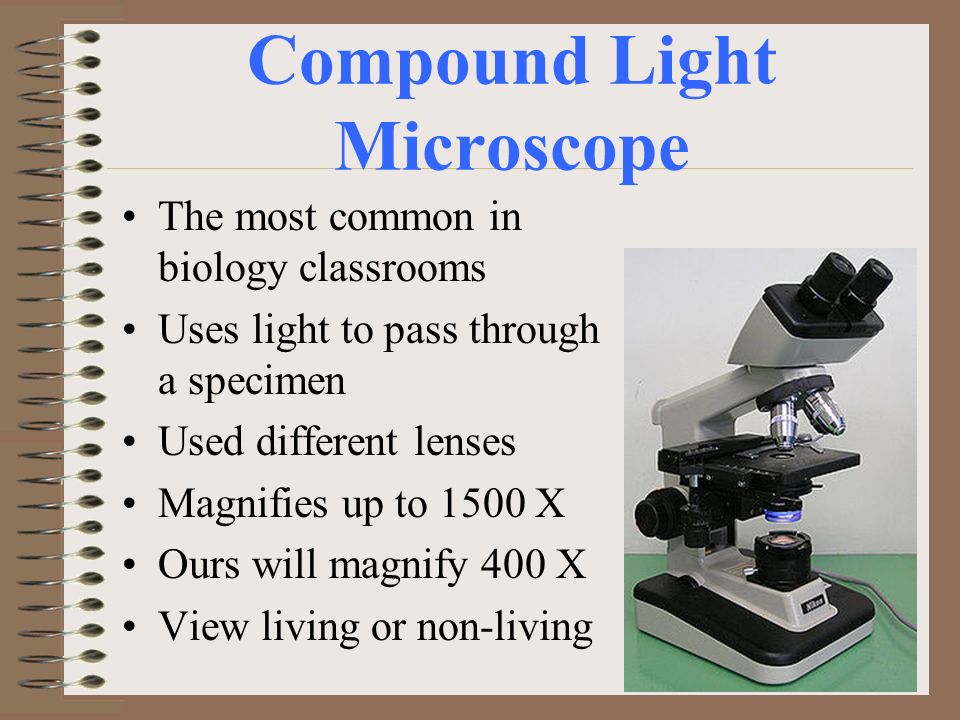
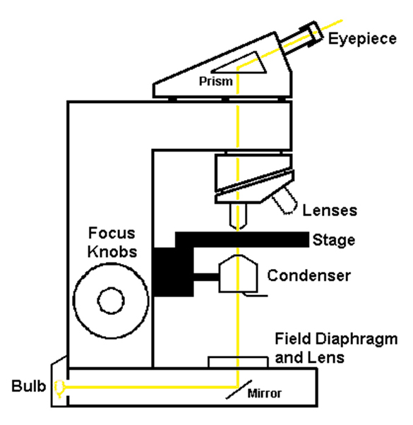
The compound microscope uses light for illumination. Some compound microscopes make use of natural light, whereas others have an illuminator attached to the base. The specimen is placed on the stage and observed through different lenses of the microscope, which have varying magnification powers. Compound Microscope Parts and Functions And with the help of the handy microscope diagram and microscope worksheet found on this page, you’ll be an expert on light microscope parts in no time. Together, these two science worksheets make a great study guide for students preparing for an upcoming parts of a compound microscope quiz or freshman biology test. 27/05/2021 · Image 17: The field diaphragm. Picture Source: olympus-lifescience.com Bottom lens/field diaphragm – it is a knob used to adjust the amount of light that gets in contact with the specimen. (5, 6, 7, and 8) How a compound microscope works/functions? Light begins at the base of the microscope coming from the source of illumination. The light microscope can extend our ability to see detail by 1000 times, so that we can see objects as small as 0.1 micrometer (um) or 100 nanometers (nm) in diameter. The transmission electron microscope extends this capability to objects as small as 0.5 nm in diameter, 1/200,000th the size of objects that are visible to the naked eye. Without
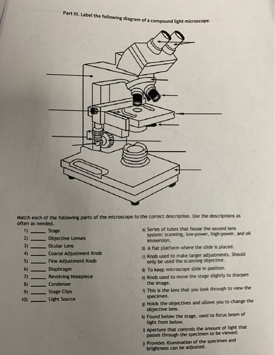
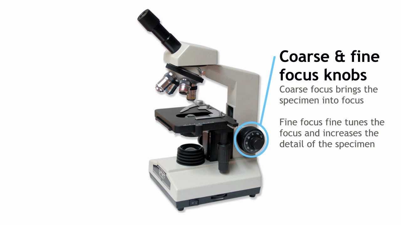
18/07/2021 · The illuminator is the light source for a microscope. A compound light microscope mostly uses a low voltage bulb as an illuminator. The stage is the flat platform where the slide is placed. Nosepiece and Aperture. Nosepiece is a rotating turret that holds the objective lenses. The viewer spins the nosepiece to select different objective lenses. Unlabelled diagram of compound microscope. This activity has been designed for use in homes and schools. Labeled microscope diagram adjusting the focus knob a compound microscopre coloring pages. The stand is made up of a heavy foot which carries a curved inclinable limb or arm bearing the body tube. Parts of a Compound Microscope Each part of the compound microscope serves its own unique function, with each being important to the function of the scope as a whole. The individual parts of a compound microscope can vary heavily depending on the configuration & applications that the scope is being used for. Common compound microscope parts include: Compound Microscope Definitions for ... A light microscope. (A) Diagram showing the light path in a compound microscope. Light is focused on the specimen by lenses in the condensor. A combination of objective lenses and eyepiece lenses are arranged to focus an image of the illuminated specimen

As light passes directly from the source to the eye through the two lenses, the field of vision is brightly illuminated. That is why; it is a bright-field microscope. Parts of a Compound Microscope: The parts of a compound microscope are of two categories as given below: (i) Mechanical Parts:

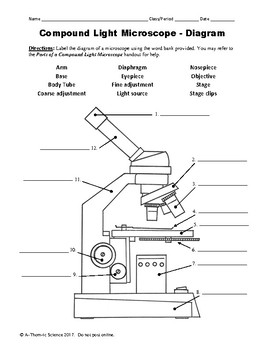
COMPOUND(LIGHT(MICROSCOPE(LAB((Follow(written(andoral(instructions.((((A.((Label(the(parts(of(the(compound(microscope.(Be(able(to(label(a(blank(diagram

Nov 4, 2021 — Learn the compound light microscope's parts and functions by viewing a compound microscope diagram. Also, read about the uses of a compound.What are the parts of the compound microscope and their functions?What are the functions of the microscope?
Compound Microscope - Diagram (Parts labelled), Principle and Uses. ... Also called as binocular microscope or compound light microscope, it is a remarkable magnification tool that employs a combination of lenses to magnify the image of a sample that is not visible to the naked eye.
For convenience, if yellow light of wave length of 580 nm with numerical aperture (NA) of 1.0 is used in the microscope, the resolution power (RP) of the microscope will be: Resolution power (RP) = 580/2 x 1 = 290 nm . Method for Studying Microbes with Compound Microscope:
Aperture. Illuminator. Condenser. Diaphragm. Parts of a Compound Microscope. Video: Parts of a compound Microscope with Diagram Explained. As a side note, the microscope used in this post is a great entry level or beginner microscope if you are trying to get someone interested in microscopes, microbiology, or science in general.

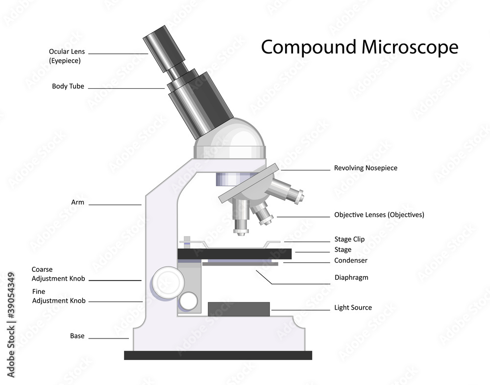
A compound light microscope is a microscope with more than one lens and its own light source. In this type of microscope, there are ocular lenses in the binocular eyepieces and objective lenses in a rotating nosepiece closer to the specimen.

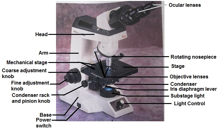
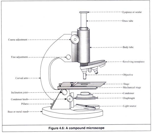
Figure: Diagram of parts of a microscope. There are three structural parts of the microscope i.e. head, base, and arm. Head - This is also known as the body, it carries the optical parts in the upper part of the microscope. Base - It acts as microscopes support. It also carries microscopic illuminators.

In the mean time we talk concerning Light Microscope Diagram Worksheet, scroll down to see particular similar images to inform you more. simple light microscope diagram, compound microscope parts blank and compound light microscope parts are three of main things we will present to you based on the gallery title.
The optical microscope, also referred to as a light microscope, is a type of microscope that commonly uses visible light and a system of lenses to generate magnified images of small objects. Optical microscopes are the oldest design of microscope and were possibly invented in their present compound form in the 17th century. Basic optical microscopes can be very simple, although many …
Compound light microscope diagram. Early microscopes like Leeuwenhoeks were called simple because they. Light used to illuminate the slide or specimen from the base of the microscope. Anatomy Of A Microscope Microscope Illumination Olympus Life. ARM Used to SUPPORT the. Structural element connects the head to the base.
Microscope Parts and Functions With Labeled Diagram and Functions How does a Compound Microscope Work?. Before exploring microscope parts and functions, you should probably understand that the compound light microscope is more complicated than just a microscope with more than one lens.. First, the purpose of a microscope is to magnify a small object or to magnify the fine details of a larger ...

A compound light microscope is a type of light microscope that uses a compound lens system, meaning, it operates through two sets of lenses to magnify the image of a specimen. It's an upright microscope that produces a two-dimensional image and has a higher magnification than a stereoscopic microscope.
All modern light microscopes are made up of a combination of more than one glass lens. The main parts of compound microscope are the condenser lens, the objective lens, and the eyepiece lens, and these instruments are referred to as compound microscopes.
A) toward A B) toward Increase the amount of light passing through the specimen. A) open the diaphragm B C) toward C D) toward D 17. The diagram below represents a cell in the field of view of a compound light microscope. In which direction should the slide be moved on the microscope stage to center the cell in the field of view? Base your answer to the following question on the diagram below ...

The most familiar type of microscope is the optical, or light, microscope, in which glass lenses are used to form the image. Optical microscopes can be simple, consisting of a single lens, or compound, consisting of several optical components in line. The hand magnifying glass can magnify about 3 to 20×. Single-lensed simple microscopes can ...

Start studying Compound Microscope Diagram. Learn vocabulary, terms, and more with flashcards, games, and other study tools.
Compound Microscope. A compound microscope uses a combination of lenses coupled with an artificial light source to magnify an object at various zoom levels to study the object. A compound microscope: Is used to view samples that are not visible to the naked eye; Uses two types of lenses - Objective and ocular lenses

Compound Microscope Diagram with Parts of The Support system. The Foot: It is the heavy metallic bottom part which supports all the other parts of the microscope. It may be oval, tripod or horseshoe shaped. Tube: It is the microscope arm's tubular, hollow component that is attached to the top half of the arm. It may be adjusted up and down ...

Simple light microscopes of the past could magnify an object to 266X as in the case of Leeuwenhoek's microscope. Modern compound light microscopes, under optimal conditions, can magnify an object from 1000X to 2000X (times) the specimens original diameter. Diagram Showing Light Traveling Through The Microscope


0 Response to "41 diagram of a compound light microscope"
Post a Comment