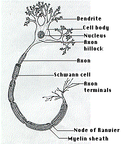
44 which part of the diagram is considered nerve fiber
Nerve conduction measures reflect the function of the best-surviving nerve fibers, and values can remain almost normal even if only a few fibers remain unaffected by the pathology. 5 Electrophysiology testing can yield up to 30% false-negative results for PN disorders. 14 Electrophysiology testing cannot differentiate intraneural pathology from extraneural nerve compression, an important point ... Which part of the diagram is considered nerve fiber? a) A b) D c) I d) Both A and D e) All of these choices make up the nerve fiber. D. This part of the neuron contains the nucleus and Nissl bodies. a) A b) B c) C d) E e) Both A and B. B.
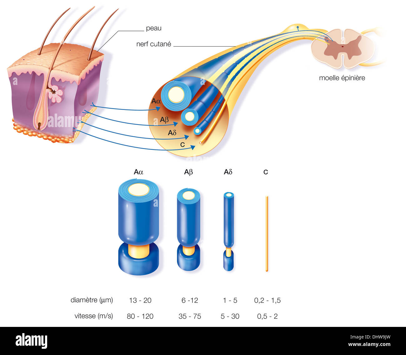
Nerve fibers connecting cortical area in one hemisphere with cortical area in ... The diagrams are based on monopolar recording of a moist nerve in air.

Which part of the diagram is considered nerve fiber
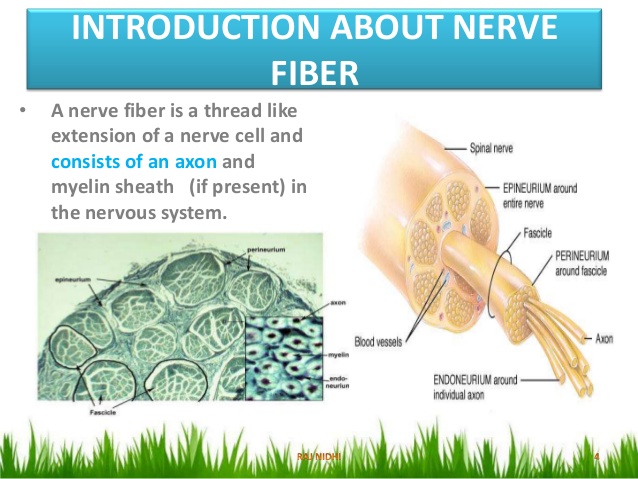
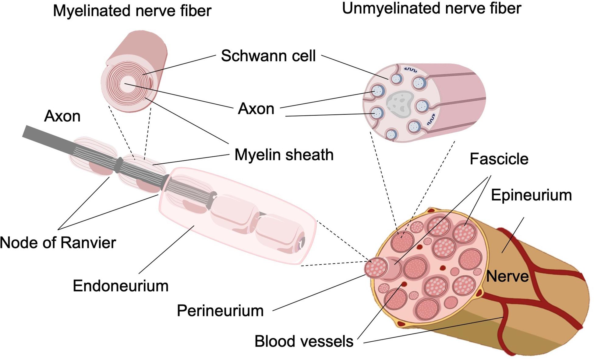
Within the endoneurium, individual nerve fibers are surrounded by a liquid called the endoneurial ... Diagram A shows the primary structures of a nerve. Which part of the diagram is considered nerve fiber? A)A B)D C)I D)Both A and D E)All of these choices make up the nerve fiber. Free. Multiple Choice . Unlock to view answer. Q 37 Q 37. This part of the neuron contains the nucleus and Nissl bodies. A)A B)B C)C D)E E)Both A and B. Free. Multiple Choice . In the standard set of conditions considered here (intact fast twitch fibers, amphibian muscle, 4 °C), v 0 is about 2 muscle lengths per second, or about 2400 nm s −1 in each half-sarcomere, a quantity that represents the relative sliding motion between each pair of apposing myosin and actin filaments. 9,25 The power output or work rate of the muscle, calculated as the load multiplied by ...
Which part of the diagram is considered nerve fiber. Cancer Definition Cancer is not just one disease, but a large group of almost 100 diseases. Its two main characteristics are uncontrolled growth of the cells in the human body and the ability of these cells to migrate from the original site and spread to distant sites. If the spread is not controlled, cancer can result in death. Description One out of ... Structure of Skeletal Muscle. A whole skeletal muscle is considered an organ of the muscular system.Each organ or muscle consists of skeletal muscle tissue, connective tissue, nerve tissue, and blood or vascular tissue.. Skeletal muscles vary considerably in size, shape, and arrangement of fibers. They range from extremely tiny strands such as the stapedium muscle of the middle ear to large ... Which part of the diagram is considered nerve fiber? a) A b) D c) I d) Both A and D e) All of these choices make up the nerve fiber. d. In the diagram, where are axon terminals? a) F b) G c) H d) I e) None of these choices. c) H. This part of the neurons contains the nissl bodies. a) A b) B c) C d) D e) both A and B. B. Which part of the diagram is considered nerve fiber? A D I Both A and D All of these choices make up the nerve fiber. Both A and D. Na+/K+-ATPase is considered to be an electrogenic pump because A. it contributes to the negativity of the resting membrane potential.
Each part of the cortex consists of the same small set of neuronal elements, laid out in a highly stereotyped geometry. At an intermediate level, the cerebellum and its auxiliary structures can be separated into several hundred or thousand independently functioning modules called "microzones" or "microcompartments". Gross anatomy. View of the cerebellum from above and behind. The cerebellum … Which part of the diagram is considered nerve fiber a a b d c i d both a and d from biology bio 226 at harold washington college city colleges of chicago. A d i both a and d all of these choices make up the nerve fiber. Which part of the diagram is considered nerve fiber. This part of the diagram contains organelles and nissl bodies. Which part of the diagram is considered nerve fiber? Cervical plexus. The nerve labeled A in the diagram arises from which plexus? C** Where is the anterior (ventral) root of a spinal nerve? Bipolar neuron. What is the structural classification of the neuron labeled B? D** Which nerve labeled in the diagram does NOT arise from any plexus? Difficulty: MediumStudy Objective 1: SO 12.2 Compare the structures and functions of neurons and neuroglia and white matter and gray matter.
This process of depolarization, repolarization, and recovery moves along a nerve fiber from neuron to neuron like a very fast wave. While an action potential is in progress, another cannot be generated under the same conditions. In unmyelinated axons (axons that are not covered by a myelin sheath), this happens in a continuous fashion because there are voltage-gated channels throughout the ... Which part of the diagram is considered nerve fiber? A, B. This part of the diagram contains organelles and Nissl bodies. E. This portion of the diagram contains cytoplasm and a myelin sheath wrapped around neurolemma. C. In the diagram, where is the axon collateral? H. An axon or nerve fiber is a long, slender projection of a nerve cell, or neuron, ... The axon hillock is the area formed from the cell body of the neuron as ... Halteres (/ h æ l ˈ t ɪər iː z /; singular halter or haltere) (from Ancient Greek: ἁλτῆρες, weights held in the hands to give an impetus in leaping) are a pair of small club-shaped organs on the body of two orders of flying insects that provide information about body rotations during flight. Examples of insects with halteres are houseflies, mosquitoes, gnats, and craneflies.
Neuro Clickers 1 Flashcards Quizlet
08.10.2011 · The ganglion cell axons run in the nerve fiber layer above the inner limiting membrane towards the optic nerve head in a arcuate form (Fig. 00, streaming pink fibers). The fovea is, of course, free of a nerve fiber layer as the inner retina and ganglion cells are pushed away to the foveal slope. The central ganglion cell fibers run around the foveal slope and sweep in the direction of the ...

Nerve Tissue Synapses And Neurotransmitters Knowledge Amboss
Which part of the diagram is considered nerve fiber. This part of the diagram contains organelles and nissl bodies. Dec 9 2013. Study 11 ch 12 diagrams for the final. The thoracic nerves refer to the cluster of nerve fibers found in the upper body particularly within the chest region. This part of the neuron contains the nucleu and nissl bodies.

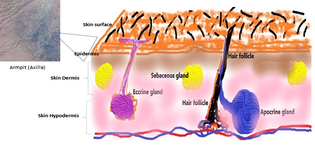
A Simplified Illustration Of The General Anatomy Of The Skin With The Download Scientific Diagram
The optic nerve is considered part of the central nervous system. The myelin on the optic nerve is produced by oligodendrocytes rather than Schwann cells and it is encased in the meningeal layers instead of the standard endoneurium, perineurium, and epineurium of the peripheral nervous system. The optic nerve travels through the optic canal, partially decussates in the optic chiasm, and ...
Nerve Fiber Drawing Stock Photo Alamy
Which part of the diagram is considered nerve fiber? all of these choices make up the nerve fiber both a and d a I d. c. remove a neurotrasmitter. Diffusion, enzymatic degradation, and uptake by cells are all ways to a. inhibit a presynaptic potential b. stop a spatial summation

Efferent Nerve Fiber An Overview Sciencedirect Topics
Which part of the diagram is considered nerve fiber. Related diagrams and images facial nerve. Refer to image 1 37. 69 the function of this pathway is to convey nerve impulses from the brainstem to cause automatic movements that regulate muscle tone posture and balance and orientation of the head and body.
Facial Nerve Paralysis Overview Anatomy Pathophysiology
28.05.2021 · A comprehensive database of more than 35 histology quizzes online, test your knowledge with histology quiz questions. Our online histology trivia quizzes can be adapted to suit your requirements for taking some of the top histology quizzes.

A Schematic Of The Nerve Fiber With Paranode B The Myelinated Axon Download Scientific Diagram
In the standard set of conditions considered here (intact fast twitch fibers, amphibian muscle, 4 °C), v 0 is about 2 muscle lengths per second, or about 2400 nm s −1 in each half-sarcomere, a quantity that represents the relative sliding motion between each pair of apposing myosin and actin filaments. 9,25 The power output or work rate of the muscle, calculated as the load multiplied by ...

Localized Retinal Nerve Fiber Layer Defects And Stroke Stroke
Which part of the diagram is considered nerve fiber? A)A B)D C)I D)Both A and D E)All of these choices make up the nerve fiber. Free. Multiple Choice . Unlock to view answer. Q 37 Q 37. This part of the neuron contains the nucleus and Nissl bodies. A)A B)B C)C D)E E)Both A and B. Free. Multiple Choice .
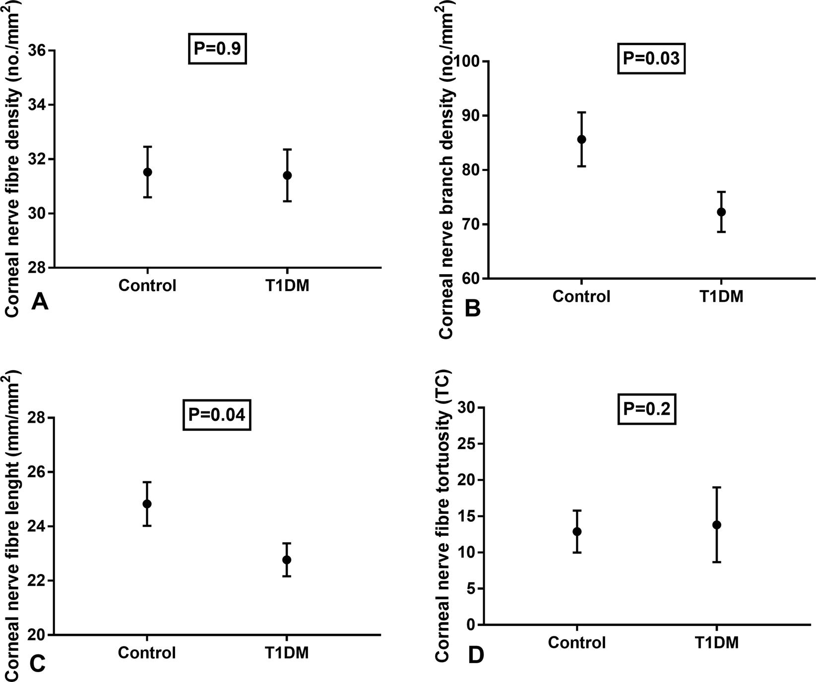
Early Corneal Nerve Fibre Damage And Increased Langerhans Cell Density In Children With Type 1 Diabetes Mellitus Scientific Reports
Within the endoneurium, individual nerve fibers are surrounded by a liquid called the endoneurial ... Diagram A shows the primary structures of a nerve.

Nerve Fiber An Overview Sciencedirect Topics

Neuro Clickers 1 Flashcards Quizlet
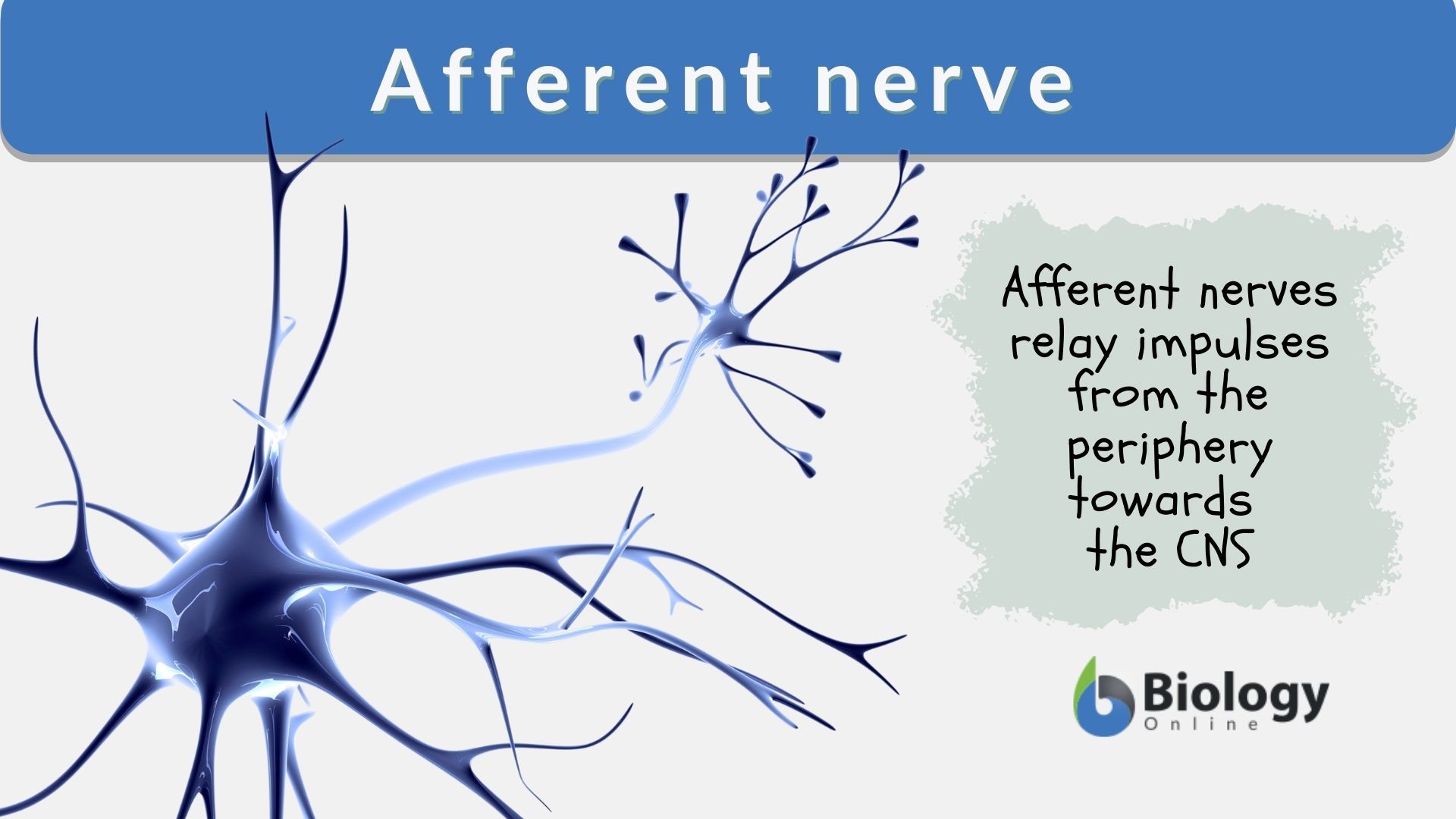
Afferent Nerve Definition And Examples Biology Online Dictionary

Chapter 12 Diagrams From Test Bank Flashcards Quizlet
.jpg)
Peripheral Nerve Fiber Types Mountain West Foot Ankle Institute

Which Part Of The Diagram Is Considered Nerve Fiber A A B D C I D Both A And D Course Hero

Synovial Nerve Fiber Density Decreases With Naturally Occurring Osteoarthritis In Horses Osteoarthritis And Cartilage

Exposure To Secondhand Smoke In Children Is Associated With A Thinner Retinal Nerve Fiber Layer The Hong Kong Children Eye Study American Journal Of Ophthalmology
Malodor Archives International Scientific Association For Probiotics And Prebiotics Isapp

Neuro Clickers 1 Flashcards Quizlet

Which Part Of The Diagram Is Considered Nerve Fiber A A B D C I D Both A And D Course Hero

Simulated Scattering Patterns For Different Artificial Nerve Fiber Download Scientific Diagram

Wide Field Trend Based Progression Analysis Of Combined Retinal Nerve Fiber Layer And Ganglion Cell Inner Plexiform Layer Thickness Ophthalmology

Parasympathetic Nervous System Wikipedia

Time Course Changes In Optic Nerve Head Blood Flow And Retinal Nerve Fiber Layer Thickness In Eyes With Open Angle Glaucoma Ophthalmology

Impact Of Artifacts From Optical Coherence Tomography Retinal Nerve Fiber Layer And Macula Scans On Detection Of Glaucoma Progression American Journal Of Ophthalmology
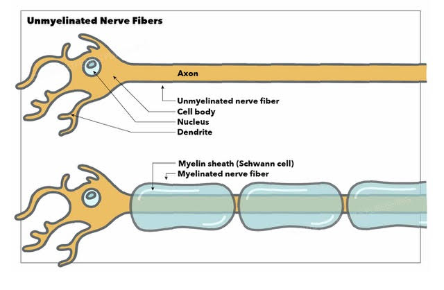
Neuroanatomy Unmyelinated Nerve Fibers Article

Which Part Of The Diagram Is Considered Nerve Fiber A A B D C I D Both A And D Course Hero
Nerve Fiber Liberal Dictionary
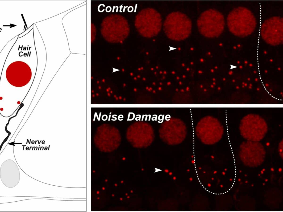
Noise Induced Hidden Hearing Loss Mechanism Nerve Fiber Loss Discussed

Ganglion Cell Inner Plexiform Layer And Retinal Nerve Fiber Layer Changes In Glaucoma Suspects Enable Prediction Of Glaucoma Development American Journal Of Ophthalmology
Frontiers Biomimetic Approaches For Separated Regeneration Of Sensory And Motor Fibers In Amputee People Necessary Conditions For Functional Integration Of Sensory Motor Prostheses With The Peripheral Nerves Bioengineering And Biotechnology
Neurons

The Anti Inflammatory Effects Of Glucagon Like Peptide Receptor Agonist Lixisenatide On The Retinal Nuclear And Nerve Fiber Layers In An Animal Model Of Early Type 2 Diabetes The American Journal Of Pathology

Intraepidermal Nerve Fibers Increase In Dry Skin Of Acetone Treated Mice Journal Of Dermatological Science

Ganglion Cell Inner Plexiform Layer And Retinal Nerve Fiber Layer Changes In Glaucoma Suspects Enable Prediction Of Glaucoma Development American Journal Of Ophthalmology

2 293 Nerve Fiber Illustrations Clip Art Istock

A Pgp9 5 Positive Nerve Fibers Brown In Direct Contact With A Bile Download Scientific Diagram
Ijms Free Full Text A Novel In Vitro Assay Using Human Ipsc Derived Sensory Neurons To Evaluate The Effects Of External Chemicals On Neuronal Morphology Possible Implications In The Prediction Of Abnormal

Ijms Free Full Text Bioactive Nanofiber Based Conduits In A Peripheral Nerve Gap Management An Animal Model Study Html

14 Chapter 13 The Spinal Cord Ideas Spinal Cord Spinal Chapter 13

Diabetic Neuropathy And The Sensory Neuron New Aspects Of Pathogenesis And Their Treatment Implications Kobayashi 2018 Journal Of Diabetes Investigation Wiley Online Library

Optical Coherence Tomography Normative Peripapillary Retinal Nerve Fiber Layer And Macular Data In Children 0 5 Years Of Age American Journal Of Ophthalmology
Facial Nerve Paralysis Overview Anatomy Pathophysiology
Plos One Bruch S Membrane Opening Minimum Rim Width And Retinal Nerve Fiber Layer Thickness In A Brazilian Population Of Healthy Subjects
0 Response to "44 which part of the diagram is considered nerve fiber"
Post a Comment