45 inner ear crystals diagram
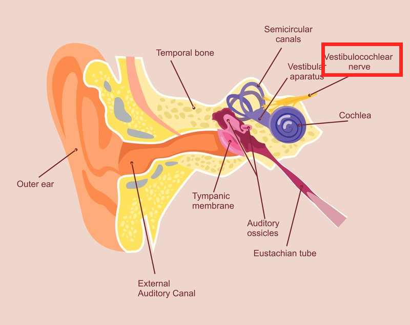
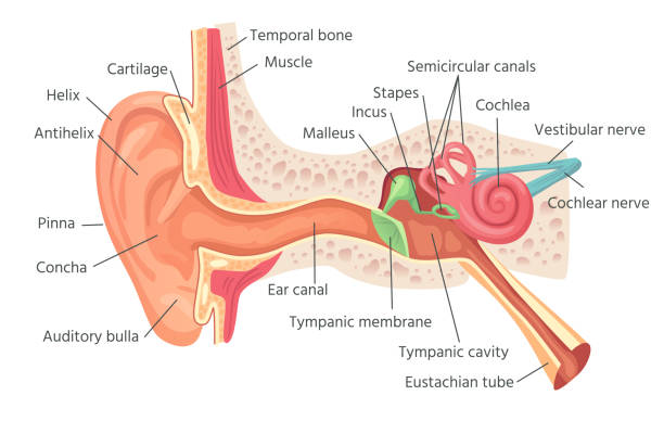
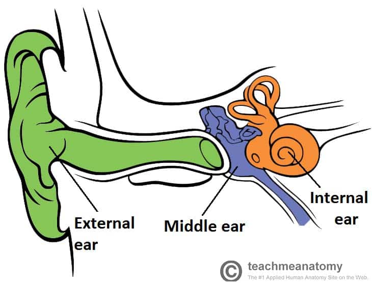
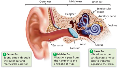
Your inner ear is the deepest part of your ear.. The inner ear has two special jobs. It changes sound waves to electrical signals (nerve impulses). This allows the brain to hear and understand sounds. The Normal Ear. Hearing and Balance. The human ear can be divided into three sections. Each section performs a different rold in transmitting sound waves to the brain. Outer ear. Middle ear. Inner ear. View the diagrams below to learn more about the different sections of the ear and how we hear.
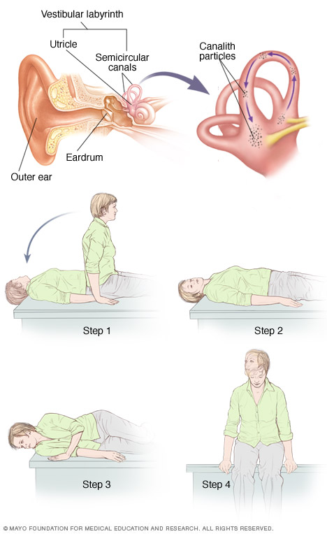
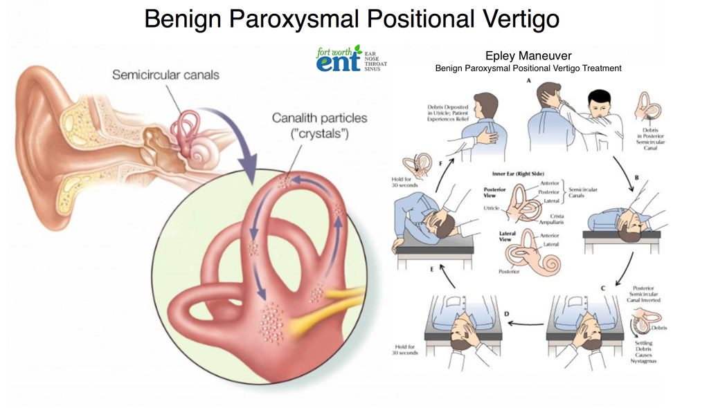
Benign Paroxysmal Positioning Vertigo is caused by loose inner ear crystals in the inner ear that migrate while sleeping to the back-bottom inner ear balance canal, the so-called "posterior semi-circular canal." The maneuver demonstrated below is the way to reposition the loose crystals so that the symptoms caused by the loose crystals go away.

Inner ear crystals diagram
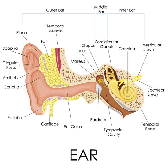
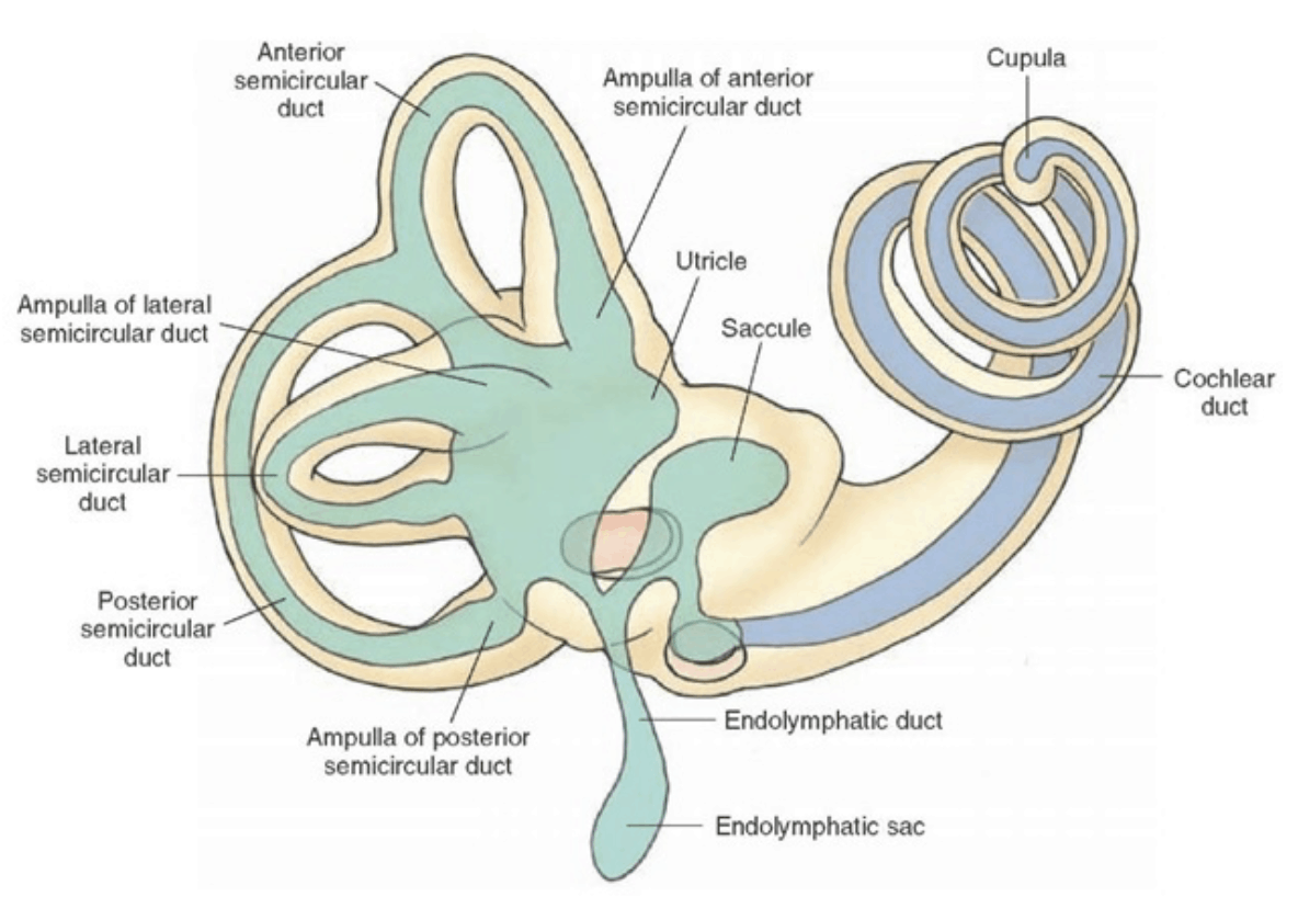
This diagram is of the outer ear. This is the part of the ear that people can see, and funnels sound into your ear canal. The rim of the pinna. A curved panel of cartridge. Part of the pinna. Bowl-shaped part of pinna. The small, hard bump above your ear lobe. Part of the pinna. ... Inner Ear - In the inner ear, vibrations are turned into ... Canalith repositioning procedure: The canalith repositioning procedure can help relieve benign paroxysmal positional vertigo (BPPV), a condition in which you have brief, but intense, episodes of dizziness that occur when you move your head. Vertigo usually comes from a problem with the part of the inner ear responsible for balance (vestibular ... Our ears are naturally lined with crystals! The inner ear plays a HUGE role in telling the brain about what our body is doing, and help us stay balanced. This part of the inner ear is formed by 3 loops (Green - picture 1) connected to a reservoir (Blue - Picture 1). Picture 1 - Semicircular Canals of the Inner Ear.
Inner ear crystals diagram. Aug 06, 2016 · BPPV is a result of tiny crystals in your inner ear being out of place. The crystals make you sensitive to gravity and help you to keep your balance. Normally, a jelly-like membrane in your ear keeps the crystals where they belong. If the ear is damaged — often by a blow to the head — the crystals can shift to another part of the ear. membrane to the oval window of the inner ear. otoliths: calcium carbonate crystals found in the utricle and saccule of the inner ear. Damage to the otoliths may lead to BPPV. oval window: oval-shaped opening from the middle ear into the inner ear. The footplate of the stapes fits into the oval window. perilymph: the fluid that fills the space ... Jan 29, 2019 · When they are dislodged, the crystals float around in the fluid area of the balance branch of the inner ear, and you will start to feel off balance. The loose crystals will start to make people feel like they are spinning and the room is spinning around them. If you are 60 or older, you are more prone to having your ear crystals dislodge. BPPV is caused by a problem with the inner ear. Calcium crystals called canaliths can end up in the semicircular canals. If these crystals become dislodged and move around, they can cause the ...
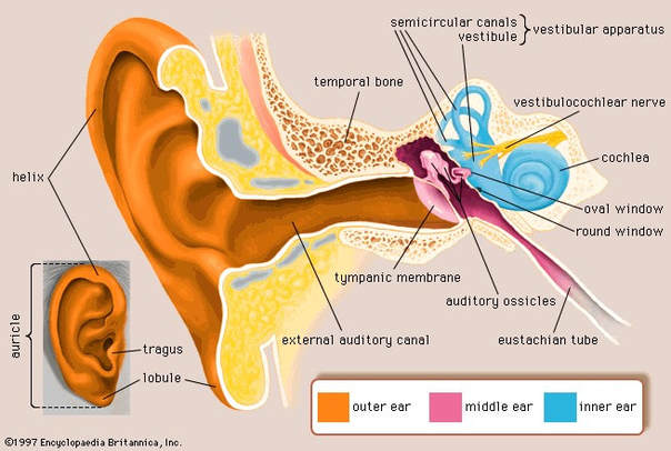
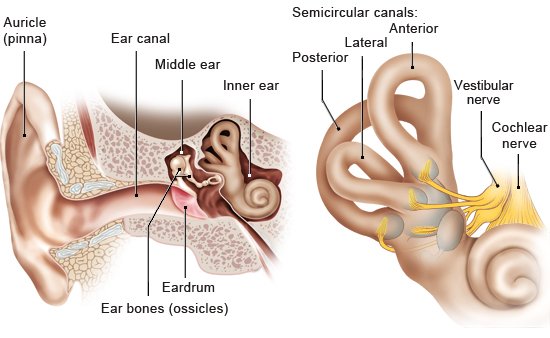
Browse 1,560 inner ear stock photos and images available, or search for inner ear illustration or inner ear diagram to find more great stock photos and pictures. Illustration of hearing, journey of the sound wave in the ear. The sound wave is captured by the auricle, penetrates in the auditory canal, vibrates... These semi-circular canals are filled with fluid and have some small calcium crystals embedded in the lining. Coming from the inner ear and running to the brain is the eighth cranial nerve, the auditory nerve. This nerve carries both balance and hearing information to the brain. Along with the eighth cranial nerve runs the seventh cranial nerve. The crystals become trapped in the inner ear's fluid-filled semicircular canal. Usually the posterior semicircular canal is affected because its structure works with the pull of gravity. The semicircular canals are normally not sensitive to head and body position changes. With BPPV, however, head and body movements such as lying down cause ... All the best Inner Ear Drawing 37+ collected on this page. Feel free to explore, study and enjoy paintings with PaintingValley.com ... Inner Ear Crystals A... 414x336 0 0. Like JPG. Inner Ear Diagram - ... 2160x2160 0 0. Like JPG. Inner Ear Stock Vide... 852x480 0 0. Like PNG. Inner Ear - Inner Ea... 450x325 0 0. Like JPG. Inner Ear Anatomy Ar...
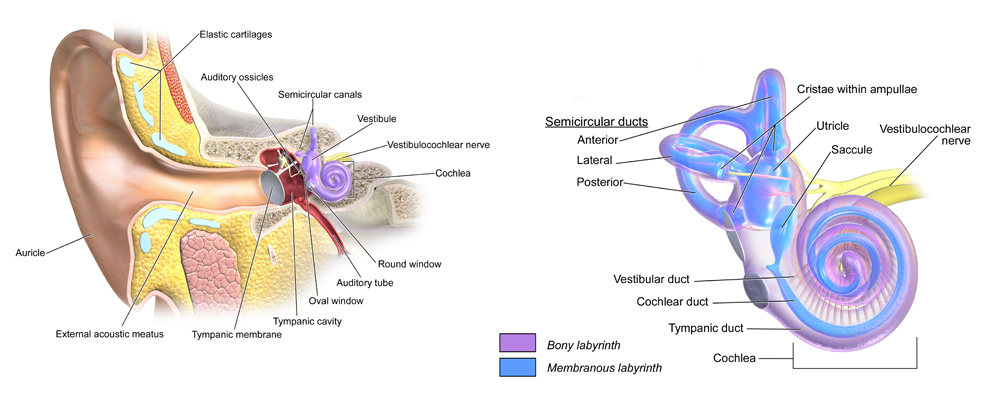
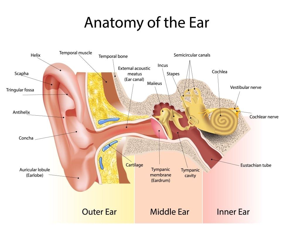
Jul 10, 2021 · The utricle is one of two "otolithic organs" in the human ear, the utricle and saccule. On the diagram above, the utricle are located in the vestibule, between the semicircular canals (5), and the cochlea (9). ... Rather than having many crystals as is the case in humans, each "otolith" in fish is made of one large crystal. ... Josephson, R ... It happens when small crystals of calcium get loose in your inner ear. You may feel it when you're getting in or out of bed, or tilting your head up. People over age 60 are more likely to get BPPV. Inner Ear 'Rock Slides' Lead To Vertigo Tiny crystals, or ear rocks, in the inner ear help us to balance. But when these pebbles fall into the sensitive ear canal, they can cause dizziness. Human ear. The ear is divided into three anatomical regions: the external ear, the middle ear, and the internal ear (Figure 2). The external ear is the visible portion of the ear, and it collects and directs sound waves to the eardrum.
Benign Paroxysmal Positional Vertigo Bppv Treatment Tests Symptoms
Disease explanation with otoconia crystals disease comparison diagram Inner Ear stock illustrations. Ear - Anatomy Frontal section through the external, middle, and internal ear. Inner Ear stock illustrations. Wireframe of the internal structure of the human ear. 3D. Front view. Vector illustration Wireframe of the internal structure of the ...
The Anatomy Of Vertigo Part Iii Of My Anatomy Of Series If By Dr Jonathan Chung Medium
One of the best and most effective options in treating vertigo is Half Somersault maneuver that is an exercise designed to bring relief by restoring the otoconia (inner ear crystals) to their natural position. You will learn how to prepare for and perform the maneuver. Also, you'll find a helpful video which guides you step by step so you can be sure you're doing everything correctly.

Inner Ear Rock Slides Lead To Vertigo Npr
Blame it on crystals. BPPV happens when tiny crystals of calcium carbonate in one part of your inner ear become dislodged and float into another part. That doesn't sound too serious, but small head movements cause the loose crystals to move, triggering your inner-ear sensors to send mixed messages to your brain.
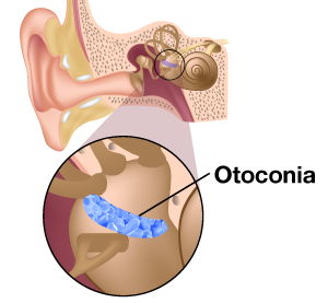
Otoconia
Nov 12, 2018 · The blades of grass represent cilia, hair-like processes that are attached to tiny nerves in your inner ear. When the crystals move, it stimulates the nerves to fire, which tells the brain your ...

Vertigo Imbalance Dizziness Synergex Physical Therapy
The inner ear has three semicircular canals which are interconnected by ×uid pathways and have gravity sensors in them. These are being capped by bed of crystals which may be dislodged from where they are and enter into any of the canals or other parts of the ear. When the dislodgement happens and crystals get into the semicircular canals, a

Ear Crystals What Are They And Their Relation To Vertigo Upper Cervical Awareness
In BPPV, these otoconia crystals escape this otolyth region and leak into one of the three semicircular ducts of the inner ear (c, e, g in diagram). These round ducts also contain a fluid. When the crystals are in this fluid, they produce motion within the fluid when the head is moved. This fluid and crystal motion excites nerve endings in the ...

Can Loose Crystals Cause Dizziness Ear And Balance
2. Wriggle your jaw. This very simple maneuver is known as the first technique of the Edmonds maneuver. Simply jut your jaw forward, then wriggle it back and forth, from side to side. If the ear blockage is mild, this action can pop your Eustachian tube open and reestablish normal air flow. Advertisement.
As The World Turns Mayo Clinic Health System
BIO MEDICAL INSTRUMENTATION. Enter the email address you signed up with and we'll email you a reset link.

Anatomy Of The Left Inner Ear Showing Displaced Otoconia From The Download Scientific Diagram
Ear Anatomy | Inside the ear | 3D Human Ear animation video | Biology | Elearnin Ear is that part of the human body that detects sound from the environment a...
Epublications Marquette Edu
Mill Hill 18002 Midnight Glass Pony Beads - Size 8/0. Leading supplier of Swarovski crystals, TOHO seed beads, Miyuki, and Czech glass beads. Nov 10, 2018 · Measuring Earrings Diameter (size) An earring can be measured by its 2 diameters: inner diameter and outer diameter (since earring is made of wire which has some thickness).

Bppv Notes From E Cervoni Md
Inner ear and balance. Loop-shaped canals in your inner ear contain fluid and fine, hairlike sensors that help you keep your balance. At the base of the canals are the utricle and saccule, each containing a patch of sensory hair cells. Within these cells are tiny particles (otoconia) that help monitor the position of your head in relation to ...

Vertigo Vector Illustration Labeled Medical Vestibular Ear Problem Scheme Anatomical Inner Earlobe And Canal Detailed Structure Disease Explanation With Otoconia Crystals Disease Comparison Diagram Royalty Free Cliparts Vectors And Stock
Crystals (otoconia) are made of calcium, and they'll shift from either one or both of the otolith organs of the inner ear. From there, they fall into one of the semicircular canals, disrupting the flow of fluid in that canal .

Vertigo Doctors Australia
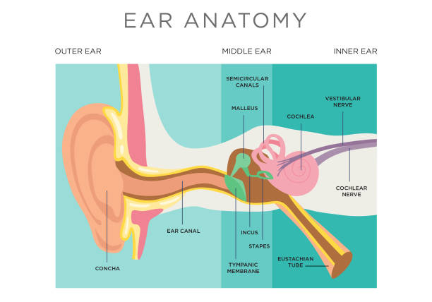
Structure and Functions of the Ear Explicated With Diagrams. The ear is another extraordinary organ of the house of wonders, that is, the human body. The ear catches sound waves and converts it into impulses, that the brain interprets, making it understandable and helps the human body differentiate between different sounds.
Bppv Overview Chester County Vertigo And Balance
The utricle is a nearby part of the ear. It contains calcium crystals (canaliths) that help it detect movement. Sometimes these crystals detach from the utricle and end up inside the semicircular canals. When these crystals move inside the canals, they may send incorrect signals to your brain about your position.

Benign Paroxysmal Positional Vertigo Emergency Care Institute Eci
Ear crystals (otolith or otoconia) are tiny calcium carbonate/calcite crystals embedded in the gelatinous otolithic membrane in the inner ear. The otolithic organs (the utricle and the saccule) are what enables you to discern which way is up even when your eyes are closed.
Bppv Baltimore Md Owings Mills Md Westminster Md Ent
Our ears are naturally lined with crystals! The inner ear plays a HUGE role in telling the brain about what our body is doing, and help us stay balanced. This part of the inner ear is formed by 3 loops (Green - picture 1) connected to a reservoir (Blue - Picture 1). Picture 1 - Semicircular Canals of the Inner Ear.

Woman 37 Has Vertigo For Four Days After Standing Near A Cannon At A Civil War Re Enactment Express Digest
Canalith repositioning procedure: The canalith repositioning procedure can help relieve benign paroxysmal positional vertigo (BPPV), a condition in which you have brief, but intense, episodes of dizziness that occur when you move your head. Vertigo usually comes from a problem with the part of the inner ear responsible for balance (vestibular ...

Vertigo Dizziness Bppv Treatment Ear Issues Perth Star Physio
This diagram is of the outer ear. This is the part of the ear that people can see, and funnels sound into your ear canal. The rim of the pinna. A curved panel of cartridge. Part of the pinna. Bowl-shaped part of pinna. The small, hard bump above your ear lobe. Part of the pinna. ... Inner Ear - In the inner ear, vibrations are turned into ...

Bppv Causes What Are They Vertigo Detective

When That Dizziness Turns Out To Be Vertigo The Washington Post
5 479 Inner Ear Stock Photos Pictures Royalty Free Images Istock
Dizziness Treatment Vertigo Epley Maneuver Fort Worth Ent

Bppv Benign Paroxysmal Positional Vertigo
Peripheral Vestibular System Avora Health

Inner Ear Rock Slides Lead To Vertigo Vertigo Treatment Vertigo Inner Ear

Supercomputing The Secrets Of The Inner Ear

What S In Your Ears Besides Wax Illinois Science Council

Vestibular Problems Blissful Balance Physiotherapy
6 Poorly Understood Conditions That Lead To Hearing Loss And Balance Problems

Inner Ear Anatomy Function And Health
The Inner Ear Bony Labyrinth Membranous Labryinth Teachmeanatomy

Spinning World Benign Paroxysmal Positional Vertigo E N T Care And Cure

Drawing Of The Left Inner Ear From Scyliorhinus Canicula Lateral View Download Scientific Diagram
Vestibular Rehabilitation Therapy Christow Canonteign Physio

When To Know Your Symptoms Of Dizziness Is Vertigo Or Bppv Orange Coast Ent Head And Neck Surgery Head And Neck Surgeons

Benign Paroxysmal Positional Vertigo Bppv Veda
How Does Our Sense Of Balance Work Informedhealth Org

Vertigo Bppv Enough To Make Your Head Spin Bellefleur Physio

159 Inner Ear Diagram Stock Photos Pictures Royalty Free Images Istock

Ali Bani Oraba Video Presentation Anatomy Of The Ear And Physiology Mechanism Of Hearing And Equilibrium

Anatomy Of The Otoliths
572 Ear Diagram Stock Photos Pictures Royalty Free Images Istock

A Medical Illustration Of Benign Paroxsymal Positional Vertigo Bppv And The Canalith Repositioning Procedure Dizziness Causes How To Cure Vertigo Vertigo
Donald Physiotherapy Blog Donald Physiotherapy
Cfzkh63qcx4w1m
0 Response to "45 inner ear crystals diagram"
Post a Comment