41 belly button anatomy diagram
The part of the umbilical arteries closest to the belly button degenerates into ligaments that serve no real purpose but the more internal part becomes part of the circulatory system and is found ... The abdomen (commonly called the belly) is the body space between the thorax (chest) and pelvis. The diaphragm forms the upper surface of the abdomen. At the level of the pelvic bones, the abdomen ...
Basically, we cut the person in a straight, vertical line from the head through the belly button and down to the toes. The median plane, therefore, creates equal right and left halves of our body.

Belly button anatomy diagram
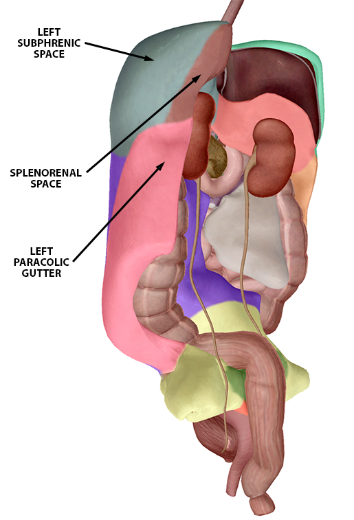
Spleen Location. Spleen is the biggest lymphoid organ present in the upper far left portion of the abdomen in the left hypochondrium and is surrounded by peritoneum. Spleen is 1 inch thick, 3 inches broad and 5 inches long. The enlargement of spleen is referred to as splenomegaly. Picture : Spleen Location and Anatomy. The real shape of your midsection boils down to a formula that includes factors like body type, fat composition, and possibly even the shape of the pelvic bone, where your ab muscles attach, says Carrie McCulloch, M.D., a musculoskeletal anatomy expert and the medical director for Kinected Pilates studio in New York City.Theoretically, a wider pelvis can translate into a broad lower abdomen ... The anatomy of your abdominal wall has many parts. Each serves an important function to give your abdomen the structure, strength, and protection it needs to look fit and work properly. Understanding the anatomy of your abdominal wall can benefit patients who are considering a tummy tuck or mummy makeover , and also women who want to to treat ...
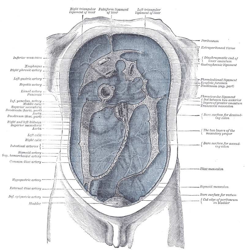
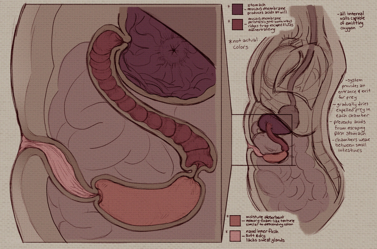
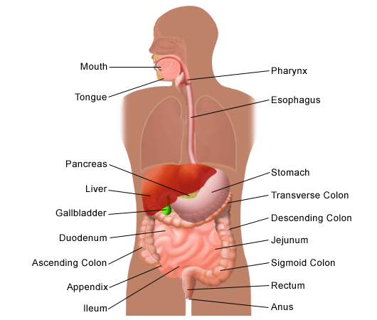
Belly button anatomy diagram. Behind your belly button there lies the Pechoti gland which houses more than 72,000 veins and millions of nerves. This has been used for centuries in other cultures as a delivery for medicine and treatments. According to those that practice this method it can be used for many an ailment. Suffering from period pain? - add a few drops of brandy ... When a person is feeling very aroused, the uterus, (and with it the cervix) moves forward towards the belly button, and very top or back of the vagina is then exposed. This area at the top or 'deep' in the vagina (shown in the illustration) is usually covered by the cervix, has a dense patch of nerves that can be very pleasurable and intense. Navel Noms Anatomy Chart. By. VoraciousPanda. 89 Favourites. 28 Comments. 11K Views. anatomy belly button chambered chart diagram digestive endo fiction guts illustration internal multi navel nom noms organs. science scientific soft speculative stomach umbilicus up vore weird endosoma made system. More. The abdominal cavity is the part of the body that houses the stomach, liver, pancreas, kidneys, gallbladder, spleen, and the large and small intestines.The diaphragm marks the top of the abdomen and the horizontal line at the level of the top of the pelvis marks the bottom. Connective tissue called the mesentery holds the abdominal organs together.
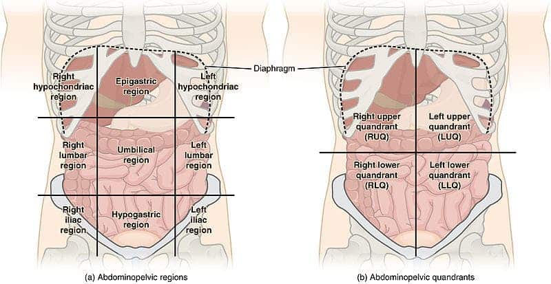
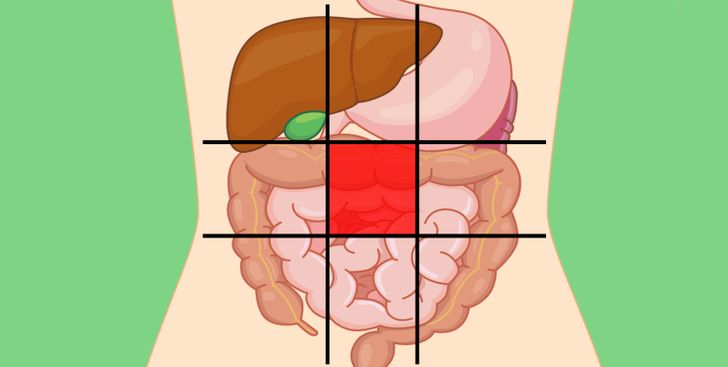
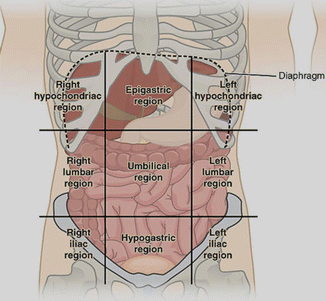
WebMD's Colon Anatomy Page provides a detailed image and definition of the colon. Learn about its function, location, and conditions that affect the colon. **UPDATE** This video got a ligament of the human body named after me! Click this link to go watch the video explaining how THAT happened... https://youtu.be... Sep 05, 2021 · The abdominopelvic quadrants can be visualized by creating two imaginary horizontal and vertical lines which intersect at the umbilicus (belly button). Four quadrants are created by the ... navel, in anatomy, a small depression in the abdominal wall at the point of attachment of the umbilical cord (q.v.). It indicates the point through which the mammalian fetus obtained nourishment from its mother through the blood vessels of the umbilical
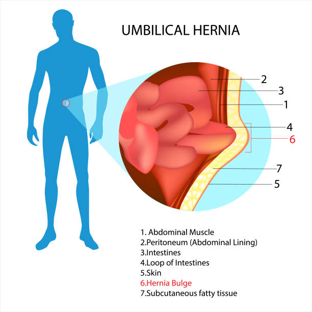
Umbilical Hernia: This hernia is one of the types of abdominal hernia that occur in the belly button. Umbilical hernia arises when a part of a small bowel pushes the abdominal wall which is very close to the navel or the belly button. Some associated conditions for these types of hernia include increased intra-abdominal pressure due to obesity, pregnancies and abdominal tumors which result in ... The Anatomy Of Your Abdominal Muscles. When you think of abs, what muscle do you typically think of? This might sound like a strange question, right? I mean, the abs are the muscle. You go to the gym to train your abs. But in actuality there are 4 separate muscles that contribute to your overall abdominal development. Navel anatomy. Navel also called umbilicus plural umbilici or umbilicuses in anatomy a small depression in the abdominal wall at the point of attachment of the umbilical cord qv. In fact the suitable anatomy for this piercing is seen much less often than its counterpart. Some navels present in the form of a more elongate hollow parallel with. Sep 07, 2021 · The systemic circuit is a part of the circulatory system that delivers blood to the organs and tissues and then returns it to the heart. Dive deeper into the definition and blood flow of the ...
Pelvis. The pelvic region is the area between the trunk — or main body — and the lower extremities, or legs. The male pelvis is different from a female's. The pelvic bones are smaller and ...

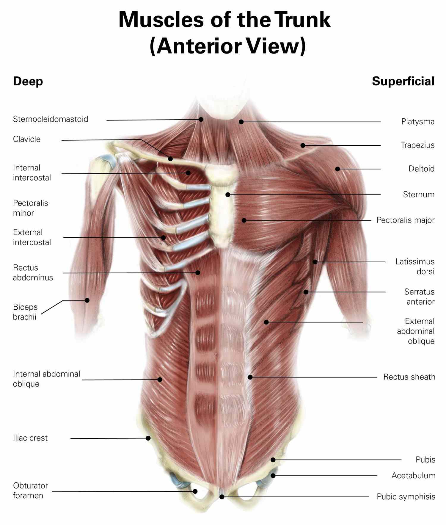
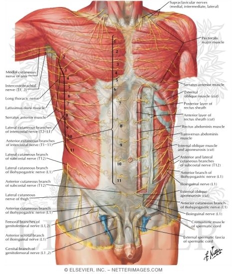
The muscles of the abdomen protect vital organs underneath and provide structure for the spine. These muscles help the body bend at the waist. The major muscles of the abdomen include the rectus ...

5,373 points • 399 comments - The "Belly Button" Way To Tell If A Person Is A Male Or A Female - 9GAG has the best funny pics, gifs, videos, gaming, anime, manga, movie, tv, cosplay, sport, food, memes, cute, fail, wtf photos on the internet!

Answer (1 of 4): The shrivelled umbilical arteries and veins. These can reopen in cases of liver blockage (such as cirrhosis of the liver) when portal venous blood tries to find alternate routes back to the heart
Human Anatomy Fundamentals: Advanced Body Proportions. This post is part of a series called Human Anatomy Fundamentals. In our last session we learned the basic, generic proportions and joint alignments of the human figure, and if you've been practicing you should be ready for some diversity. The most obvious differentiation may be between men ...
The umbilicus (belly button) is also connected to other things as well, such as the falciform ligament which runs to the liver and the medial umbilical ligaments as well. Like most things that attach to the belly button, these structures were useful in utero but aren't anymore.
The navel (clinically known as the umbilicus, commonly known as the belly button) is a protruding, flat, or hollowed area on the abdomen at the attachment site of the umbilical cord. All placental mammals have a navel. Structure. The navel is the centre of the circle in this drawing of the ...

Step 2 - The incision. A panniculectomy requires a horizontally-oriented incision in the area between the pubic hairline and belly button. The shape and length of the incision will be determined by the amount of excess skin. In some instances, a vertical midline incision is necessary in individuals who have excess skin and tissue in the ...

Fasciae and ligaments of the abdominal wall Author: Onome Okpe • Reviewer: Latitia Kench Last reviewed: September 30, 2021 Reading time: 5 minutes The abdomen is the region of the trunk between the thorax and the pelvis.It is a flexible dynamic container, housing most of the organs of the digestive system and part of the urogenital system.. Those structures are contained in its cavity, the ...

Umbilical Hernia Anatomy. The CT image below shows a cross section of a patient. You can see a clear hole in the muscle at the level of the belly button, or umbilicus. This hole is considered an umbilical hernia. The image below shows a normal umbilicus with no evidence of a hole, or a hernia.
Start studying Anatomy Diagram. Learn vocabulary, terms, and more with flashcards, games, and other study tools.
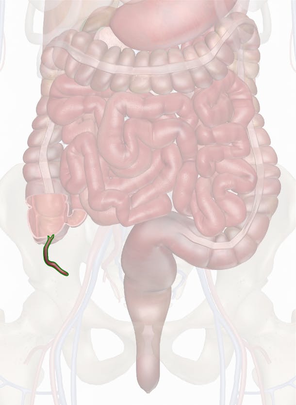
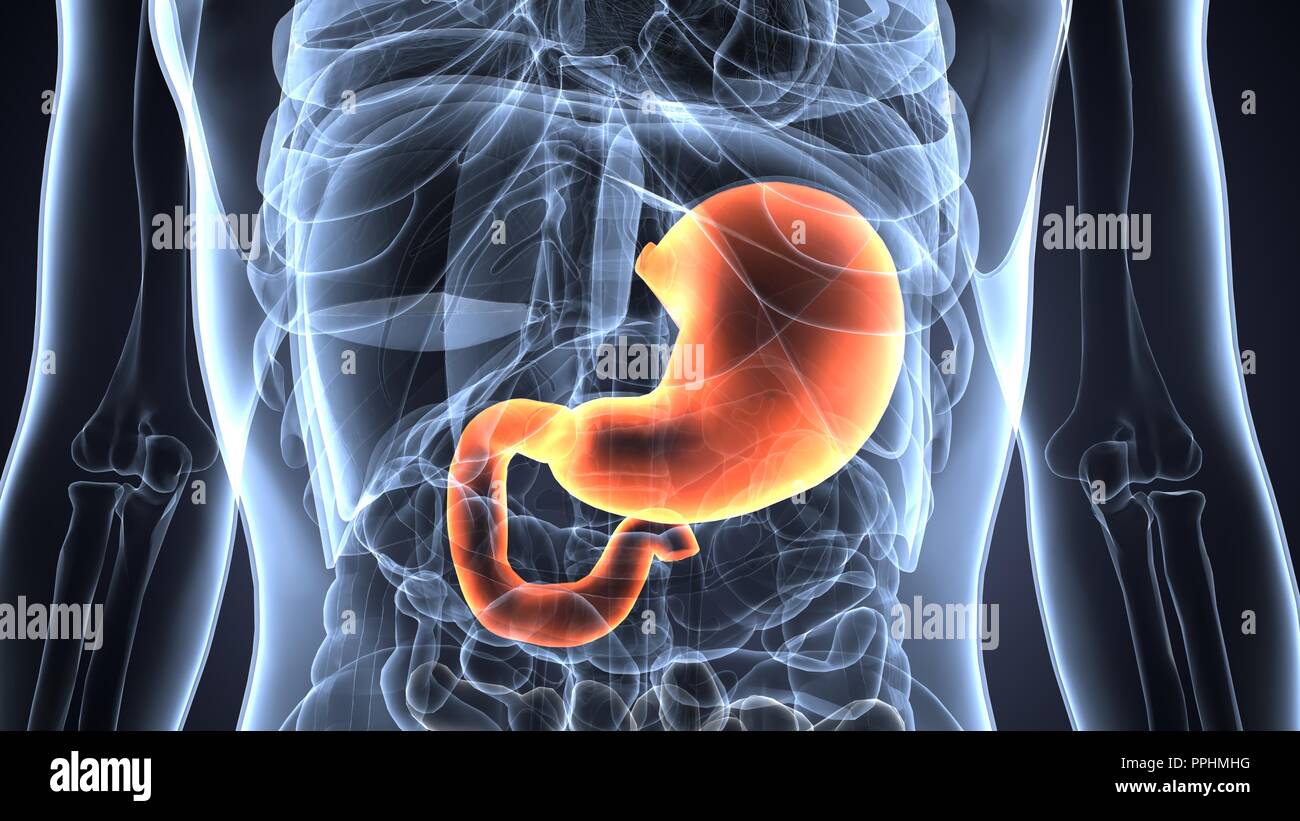
Right behind your belly button is your small intestine and part of your large intestine. Below your belly button to the right of your abdomen is your appendix. Above your belly button are your pancreas, gallbladder, and stomach. 1. Any one of these organs around the belly button could cause varying degrees of pain if they become inflamed or ...
Download Submission. Category. Artwork (Digital) / Vore. Species Unspecified / Any. Gender Other / Not Specified. Size 1280 x 845. weird made up anatomy science fiction speculative navel belly button soft vore umbilicus multi chambered internal organs chart diagram scientific illustration digestive system endo endosoma nom noms guts stomach. ★.
The anatomy of your abdominal wall has many parts. Each serves an important function to give your abdomen the structure, strength, and protection it needs to look fit and work properly. Understanding the anatomy of your abdominal wall can benefit patients who are considering a tummy tuck or mummy makeover , and also women who want to to treat ...

The real shape of your midsection boils down to a formula that includes factors like body type, fat composition, and possibly even the shape of the pelvic bone, where your ab muscles attach, says Carrie McCulloch, M.D., a musculoskeletal anatomy expert and the medical director for Kinected Pilates studio in New York City.Theoretically, a wider pelvis can translate into a broad lower abdomen ...

Spleen Location. Spleen is the biggest lymphoid organ present in the upper far left portion of the abdomen in the left hypochondrium and is surrounded by peritoneum. Spleen is 1 inch thick, 3 inches broad and 5 inches long. The enlargement of spleen is referred to as splenomegaly. Picture : Spleen Location and Anatomy.


0 Response to "41 belly button anatomy diagram"
Post a Comment