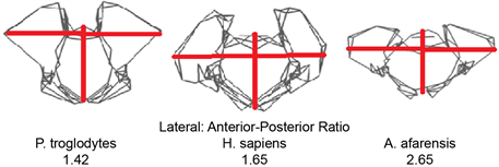
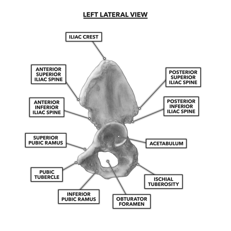
40 figure 5 11 is a diagram of the articulated pelvis
identify the bones of the shoulder and pelvic girdles. basketball career game / most powerful anti tank rifle / identify the bones of the shoulder and pelvic girdles. January 13, 2022January 13, 2022. engineering toys for 12 year olds ... he appendicular skeleton (see Figure 7.1 on page 50) is composed of the 126 bones of the appendages and the pectoral and pelvic girdles, which attach the ...14 pages
Figure 11. FG018458. ... Use points marked by arrows in diagram when getting on or off machine. Do not carry tools or supplies when you mount or dismount the machine. ... Figure 5. Operating ...

Figure 5 11 is a diagram of the articulated pelvis
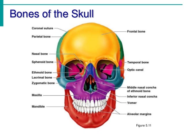
Free Printable Bones Of The Human Body Figure 1 1 The Human Skeleton Anterior View Skeletal System Worksheet Human Skeleton Anatomy Skeletal System. Blank Skeleton Diagram To Label Front And Back Of The Outstanding Skeletal System Anatomy Human Skeletal System Human Skeleton Labeled. Learning About Bones Activities For Kids And Free Skeleton ... Identify all indicated bones (or groups of bones) in the diagram of the articulated skeleton below. Parietal. Temporal. Occipital. Sternum forsendal.10 pages Label the main bones for your body printable. Find the clavula sternum scapula skull rib humerus backbone tibia os coxae radius femur ulna patella fibula and foot bones. Unlabeled pelvis bone anatomy skeletal system diagram worksheet and human skeleton bones worksheet are some main things we will show you based on the post title.
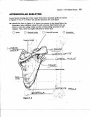
Figure 5 11 is a diagram of the articulated pelvis. Nov 24, 2021 · Figure 511 is a diagram of the articulated pelvis. The large bones of the lower limbs support the trunk when standing. The bones comprising the wrist include the distal ends of the radius and ulna 8 carpal bones and the proximal portions of the 5 metacarpal bones labeled diagram of the skeletal system pic stock pictures royalty-free photos images. The pelvic inlet involves three of the four units of which the bone pelvis is composed. The pelvic brim involves the first sacral segment, the iliac and pubis portion, but not the ischium. The pelvic inlet is delineated by a bone crest that defines its limit (the pelvic brim), which later refers to the promontory of the sacrum. Figure 5-11 is a diagram of the articulated pelvis. Select different colors for the structures listed below and use them to color the coding circles and the corresponding structures in the figure. Using the Key Choices, write the letters on the line that correspond to the bones and bone markings. Figure 5—11 is a diagram of the articulated pelvis. Identify the bones and bone markings indicated by leader lines on the figure. Select different colors for the structures listed below and use them to color the coding circles and the corresponding structures in the figure. Also, label the dashed line show-
the labels for the diagram on the left below and provide …. 6 out of 5 stars. Bones of the axial and appendicular skeleton. Blank skeleton diagram for students to complete. Name the exception. With that factor, the functions possessed by the skeleton template are also different. Human Skeleton Printables source : pinterest. 1/3 life-size two-part muscular figure. All superficial muscles of the human form are accurately reproduced in lifelike colors. The chest plate is removable to reveal the internal organs, and the right side contains a female mammary gland. There are 125 hand-numbered and identified structures of the human anatomy on this muscular figure. Figure 5-11 is a diagram of the articulated pelvis. Identify the bones and bone markings indicated by leader lines on the figure. Select different colors for the structures listed below and use them to color the coding circles and the corresponding structures in the figure. Also, label the dashed line show- ing the dimensions of the true pelvis and that showing the diameter of the false pelvis. Complete the illustration by labeling the following bone mark- ings: obturator foramen, iliac ... Figure 5-11 is a diagram of the articulated pelvis. (A) Identify the bones and bone markings indicated by leader lines on the figure.24 pages
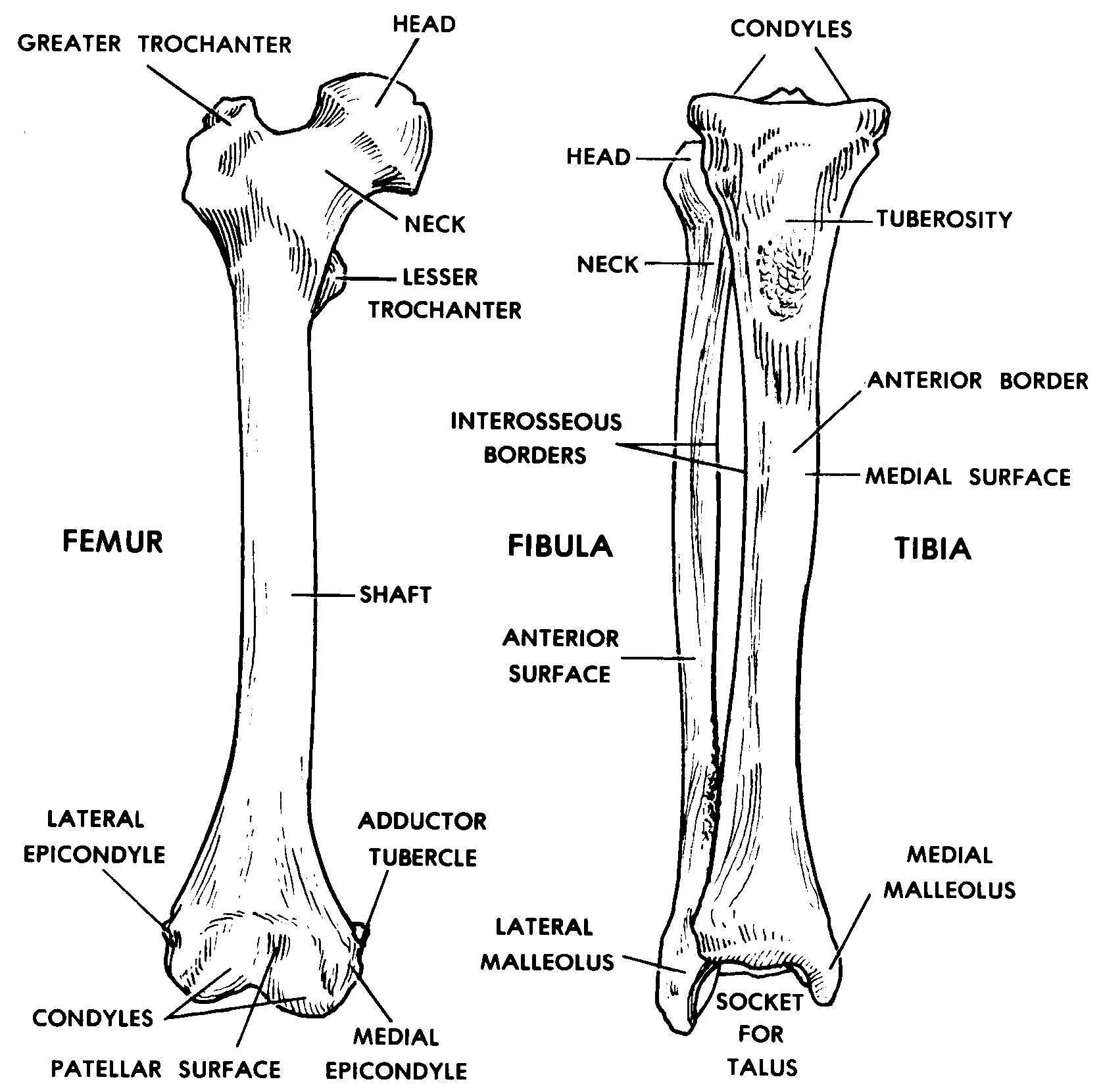
Science Skills Worksheets with low Key. Figure 5-13 is a diagram of the articulated skeleton. Appendicular skeleton worksheets bones include a lateral view of specific parts of neighboring unlabeled anatomy, answer based quiz to customize ads. Come with skull bones quiz approach and power view quiz 1 - quiz quiz. The appendicular skeleton is composed of the 126 bones of the appendages and the pectoral and pelvic girdles, which attach the limbs to the axial skeleton. 35 Blank Skull To Label. ) Dot * 1L Medium, with label 1 Medium, no label 2 Small, no label 3 Tiny, no label. Figure 5-13 is a diagram of the articulated skeleton in anatomical position Identify all. This type this article do not exist anymore the requested location in school site hierarchy. The tibia and fibula are the bones of liberty lower leg. Clinically centered; consistently and clearly illustrated; and logically organized; Gray's Atlas of Anatomy; 3rd Edition; (PDF) the companion resource to the
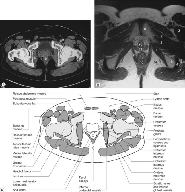
The pelvic cavity is a bowl-like structure that sits below the abdominal cavity. The true pelvis, or lesser pelvis, lies below the pelvic brim (Figure 1). This landmark begins at the level of the sacral promontory posteriorly and the pubic symphysis anteriorly. The space below contains the bladder, rectum, and part of the descending colon. In females, the pelvis also houses the uterus ...
No information is available for this page.Learn why5 pages
Figure 5—11 is a diagram of the articulated pelvis. Identify the bones and Select different colors for the structures listed below and use them to color the coding circles and the corresponding structures in the figure..
Axial skeleton diagram blank. get smaller. Appendicular Skeleton - upper limb. Clip art can be a fantastic resource and substitute for a full scale model. Click on the skeleton for a larger, printable diagram. 15% Off with code AUGUSTSTYLEZ. Skeleton printable. nnelwork, Protecbion, Levers toprouide movement ells Name the 5 categories of bones.
Pelvis - The pelvis is divided into two halves, each containing three bones, the ilium, ischium, and the pubis. Sternum and clavicle - Also known as the chest bone, the sternum is a flat bone in the chest protecting the heart and lungs, the ribs connect to the sternum via cartilage. The clavicle attaches to the sternum and shoulder blades ...
Abstract. Electromagnetic (EM) tracking has been used to quantify biomechanical parameters of the lower limb and lumbar spine during ergometer rowing to improve performance and reduce injury. Optical motion capture (OMC) is potentially better suited to measure comprehensive whole-body dynamics in rowing.
Figure 5-11 is a diagram of the articulated pelvis. Identify the bones and bone markings indicated by leader Jines on the figure, Select different colors.36 pages
The lower extremity skeleton is made up of the pelvis, hips, legs and feet. Our free textbooks. The stapes, in the middle ear, is the smallest and lightest bone of the human skeleton. Most, but not all, features you are required to know are shown on the following pages. Axial Skeleton Labeling Exercises.
This set of beginning-level materials is designed for students with no previous exposure to the Korean language. For Students 4th - 6th. Options: river, waterfall, lake, hot spring, geyser 5. Nonfiction text structures refer to the way that a text is organized. Figure 5—11 is a diagram of the articulated pelvis.
A — HUMAN NECESSITIES; A61 — MEDICAL OR VETERINARY SCIENCE; HYGIENE; A61B — DIAGNOSIS; SURGERY; IDENTIFICATION; A61B18/00 — Surgical instruments, devices or methods for tr
THE SKELETON - BLANK DIAGRAM. axial skeleton labeling quiz blank skull diagram to label awesome 7 best skeletal system images on pinterest of blank skull diagram to label {Label Gallery} Get some ideas to make labels for bottles, jars, packages, products, boxes or classroom activities for free. Transcribed Image Textfrom this Question.
Anatomy and Physiology questions and answers. 89. Chap 5 The Skal System 25. Figure 5-11 is a diagram of the articulated pelvis. Identify the bones and e markings indicated by leader lines on the figure Select different colors for the structures listed below and use them to color the coding circles and the coresponding structures in the figure. Also, Label the dashed line show in the dimensions of the.
Volume 2 Part 1
Label the main bones for your body printable. Find the clavula sternum scapula skull rib humerus backbone tibia os coxae radius femur ulna patella fibula and foot bones. Unlabeled pelvis bone anatomy skeletal system diagram worksheet and human skeleton bones worksheet are some main things we will show you based on the post title.
Identify all indicated bones (or groups of bones) in the diagram of the articulated skeleton below. Parietal. Temporal. Occipital. Sternum forsendal.10 pages
Free Printable Bones Of The Human Body Figure 1 1 The Human Skeleton Anterior View Skeletal System Worksheet Human Skeleton Anatomy Skeletal System. Blank Skeleton Diagram To Label Front And Back Of The Outstanding Skeletal System Anatomy Human Skeletal System Human Skeleton Labeled. Learning About Bones Activities For Kids And Free Skeleton ...













0 Response to "40 figure 5 11 is a diagram of the articulated pelvis"
Post a Comment