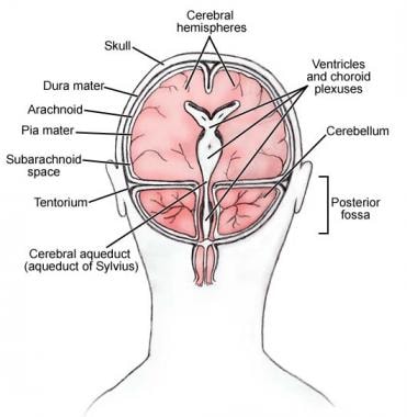
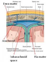
41 drag the labels onto the diagram to identify the cranial meninges and associated structures.
Drag labels onto the provided image. Sometimes a label can be used more than once, or it may not be used at all for the correct answer. When you're satisfied with your answer, select Submit.. If you can't drag one or more labels to an incorrect target, try to position the label on another target.. To clear all your labels you've placed, select Reset (next to Help). The thymus gland is a pink, lobulated lymphoid organ, located in the thoracic cavity and neck. In the adolescent, it is involved the development of the immune system. After puberty, it decreases in size and is slowly replaced by fat. Embryologically, the thymus gland is derived from the third pharyngeal pouch.
View Screen Shot 2019-02-05 at 8.26.00 PM.png from BSC 2086L at University of South Florida. Drag the labels onto the diagram to identify the cranial meninges and associated structures.
Drag the labels onto the diagram to identify the cranial meninges and associated structures.
processes visual data. Drag the labels to identify the ventricles of the brain. look at pic. Drag the labels onto the diagram to identify the cranial meninges and associated structures. look at pic. Drag the labels to identify the landmarks and features on one of the cerebral hemispheres. look at pic. Drag the labels onto the diagram to ... 1/3. This article will describe the anatomical structures which can be seen from a superior view of the skull base. This will include the various foramina, the nerves and arteries that pass through them, but also the structures of the brain and cerebellum, which all lie within the three main parts of the skull base called cranial fossae. Related for spinal cord diagram labeled label the structures on this diagram of a moss diagram labels label gallery get some ideas to make labels for bottles jars packages products boxes or classroom activities for free. Correctly identify and label the structures associated with the branches of the spinal nerve in relation to the spinal cord.
Drag the labels onto the diagram to identify the cranial meninges and associated structures.. An interactive quiz covering Spinal Cord Cross-Sectional Anatomy through multiple-choice questions and featuring the iconic GBS illustrations. Free labeling quiz. Try to understand and memorize what you can from the labeled diagram, then, try to label the cranial nerves yourself with our cranial nerves labeling quiz exercise available to download below. This is a great way to start to get the cogs turning and warm up your memory before you take our other cranial nerve quizzes (but one ... Name and describe the three meninges that cover the brain, state their functions, and locate the falx cerebri, falx cerebelli, and tentorium cerebelli. Discuss the formation, circulation, and drainage of cerebrospinal fluid. Identify the cranial nerves by number and name on a model or image, stating the origin and function of each. A sarcomere is defined as the region of a myofibril contained between two cytoskeletal structures called Z-discs (also called Z-lines), and the striated appearance of skeletal muscle fibers is due to the arrangement of the thick and thin myofilaments within each sarcomere (Figure 10.2.2).
The brain and spinal cord are enveloped within three layers of membrane collectively known as the meninges, with the cranial meninges specifically referring to the section that covers the brain. From superficial to deep, the three layers are the dura, arachnoid, and pia—the term "mater," Latin for mother, often follows these names (i.e., dura mater, arachnoid mater, pia mater).[1] The ... Drag the labels onto the diagram to identify the sel anatomy of the eye supio view Reset Help Visual axis Posterior Anterior Edge of chamber chambepupi Ciliary. LATERAL GENICULATE NUCLEUS OF THE SHEEP O 312-10h2 Largecells29 039 I I 1 mm I Fig. Download full-size image Figure 2. Chapter 15 The Autonomic Nervous System and Visceral Sensory Neurons 471 Figure 15.3). All sympathetic ganglia lie near the spinal cord and vertebral column; postganglionic axons extend from these ganglia and travel to their target organs. the . The . The cranial cavity Housed in the skull and encases the brain ... Meninges The membranes covering both the brain and the spinal cord 5 Ventral body cavity The more anterior and large of the closed body cavities; houses internal organs collectively called the viscera, or visceral organs; has two major subdivision, the thoracic cavity and the ...
Drag the labels onto the diagram to identify the cranial meninges and associated structures. 1. dura mater 2. subarachnoid space 3. pia mater 4. cerebral cortex 5. cranium 6. periosteal cranial dura 7. dural sinus 8. meningeal cranial dura 9. subdural space 10. arachnoid mater. The pelvis is a group of fused bones and may be considered the first step in the linkage of the axial skeleton (bones of the head, neck, and vertebrae) to the lower appendages. The part of the axial skeleton directly communicating with the pelvis is the lumbar spinal column. The femur is the appendicular skeletal bone connected to the pelvis at the acetabulum, a bony ring formed by the fusion ... Figure 14.2a The Spinal Cord and Spinal Meninges Anterior view of spinal cord showing meninges and spinal nerves. For this view, the dura and arachnoid membranes have been cut longitudinally and retracted (pulled aside); notice the blood vessels that run in the subarachnoid space, bound to the outer surface of the delicate pia mater. 3. On this image, the dura matter has been completely removed, you can still see the optic chiasma but the pituitary gland is missing. The infundibulum (pituitary stalk) is now visible in the center. Careful dissection also reveals two other large nerves: the oculomotor nerves (C.2). Often these two nerves are removed with the dura mater, but in this image they are still intact.
The diagram of the brain is useful for both Class 10 and 12. A Diagram Of The Parts Of The Cerebrum Brain Lobes Brain Anatomy Brain Lobes And Functions. A well-labelled diagram of a human brain is given below. Start studying Label the Brain Anatomy Diagram. Start studying Brain meninges labeled.
Directional terms describe the positions of structures relative to other structures or locations in the body. Superior or cranial - toward the head end of the body; upper (example, the hand is part of the superior extremity). Inferior or caudal - away from the head; lower (example, the foot is part of the inferior extremity).
Spinal Cord Anatomy. In adults, the spinal cord is usually 40cm long and 2cm wide. It forms a vital link between the brain and the body. The spinal cord is divided into five different parts. Several spinal nerves emerge out of each segment of the spinal cord. There are 8 pairs of cervical, 5 lumbar, 12 thoracics, 5 sacral and 1 coccygeal pair ...
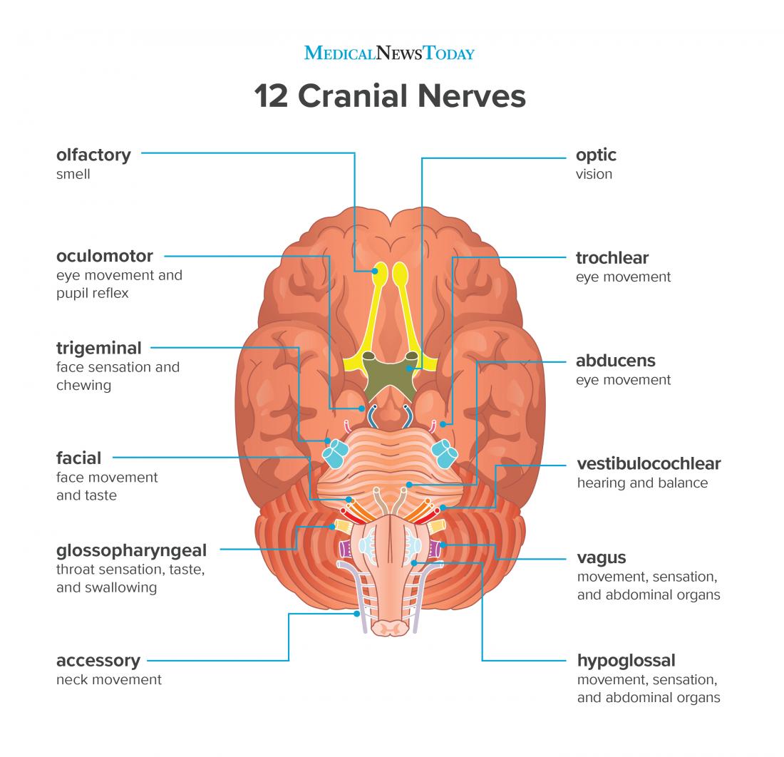
Anatomy and Physiology questions and answers. Art-labeling Activity: Origins of the Cranial Nerves (1-VI) Part A Drag the labels onto the diagram to identify the origins of the cranial nerves (1 - VI). Reset Help Abducens nerve (v Trigeminal nerve ( Olfactory tract Optic nerven Quốc Gia si Oculomotor nerve (III) Olfactory bulbs olactory nerve ...
The ventricular system is a set of communicating cavities within the brain. These structures are responsible for the production, transport and removal of cerebrospinal fluid, which bathes the central nervous system. In this article, we shall look at the functions and production of cerebrospinal fluid, and the anatomy of the ventricles that contains it.
The meninges are three connective tissue membranes that lie just external to the brain. The function of theses layers are to: 1) cover and protect the brain, 2) protect blood vessels and enclose venous sinuses, 3) contain cerebral spinal fluid, and 4) form partitions within the skull.
The cranial nerves connect through the brain stem and provide the brain with the sensory input and motor output associated with the head and neck, including most of the special senses. The major ascending and descending pathways between the spinal cord and brain, specifically the cerebrum, pass through the brain stem.
Related for spinal cord diagram labeled label the structures on this diagram of a moss diagram labels label gallery get some ideas to make labels for bottles jars packages products boxes or classroom activities for free. Correctly identify and label the structures associated with the branches of the spinal nerve in relation to the spinal cord.
1/3. This article will describe the anatomical structures which can be seen from a superior view of the skull base. This will include the various foramina, the nerves and arteries that pass through them, but also the structures of the brain and cerebellum, which all lie within the three main parts of the skull base called cranial fossae.
processes visual data. Drag the labels to identify the ventricles of the brain. look at pic. Drag the labels onto the diagram to identify the cranial meninges and associated structures. look at pic. Drag the labels to identify the landmarks and features on one of the cerebral hemispheres. look at pic. Drag the labels onto the diagram to ...




:background_color(FFFFFF):format(jpeg)/images/library/12817/anatomy-spinal-cord-cross-section_english.jpg)








0 Response to "41 drag the labels onto the diagram to identify the cranial meninges and associated structures."
Post a Comment