40 simple squamous epithelium labeled diagram
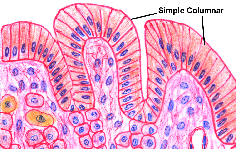
Ciliated columnar epithelium is composed of simple columnar epithelial cells with cilia on their apical surfaces. These epithelial cells ... When classifying a stratified epithelial sheet, the sheet is named for the shape of the cells in its most superficial layers. So a stratified squamous epithelium only necessarily has squamous-shaped cells in its highest layers and might have a different-shaped cell in its lower layers.
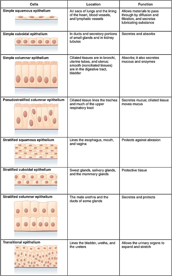
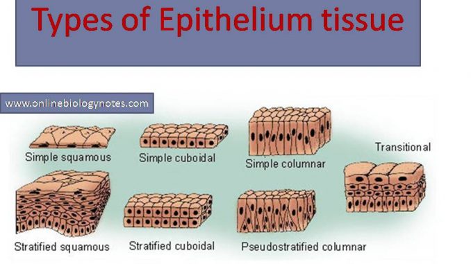
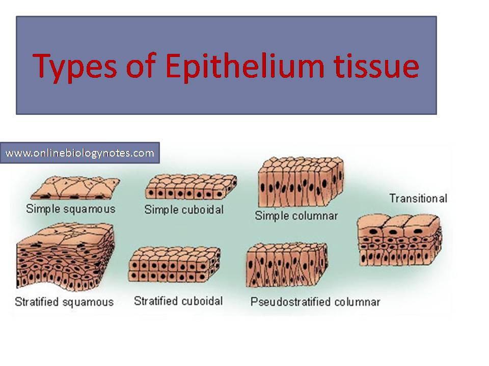
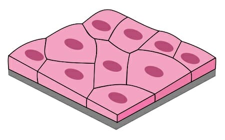
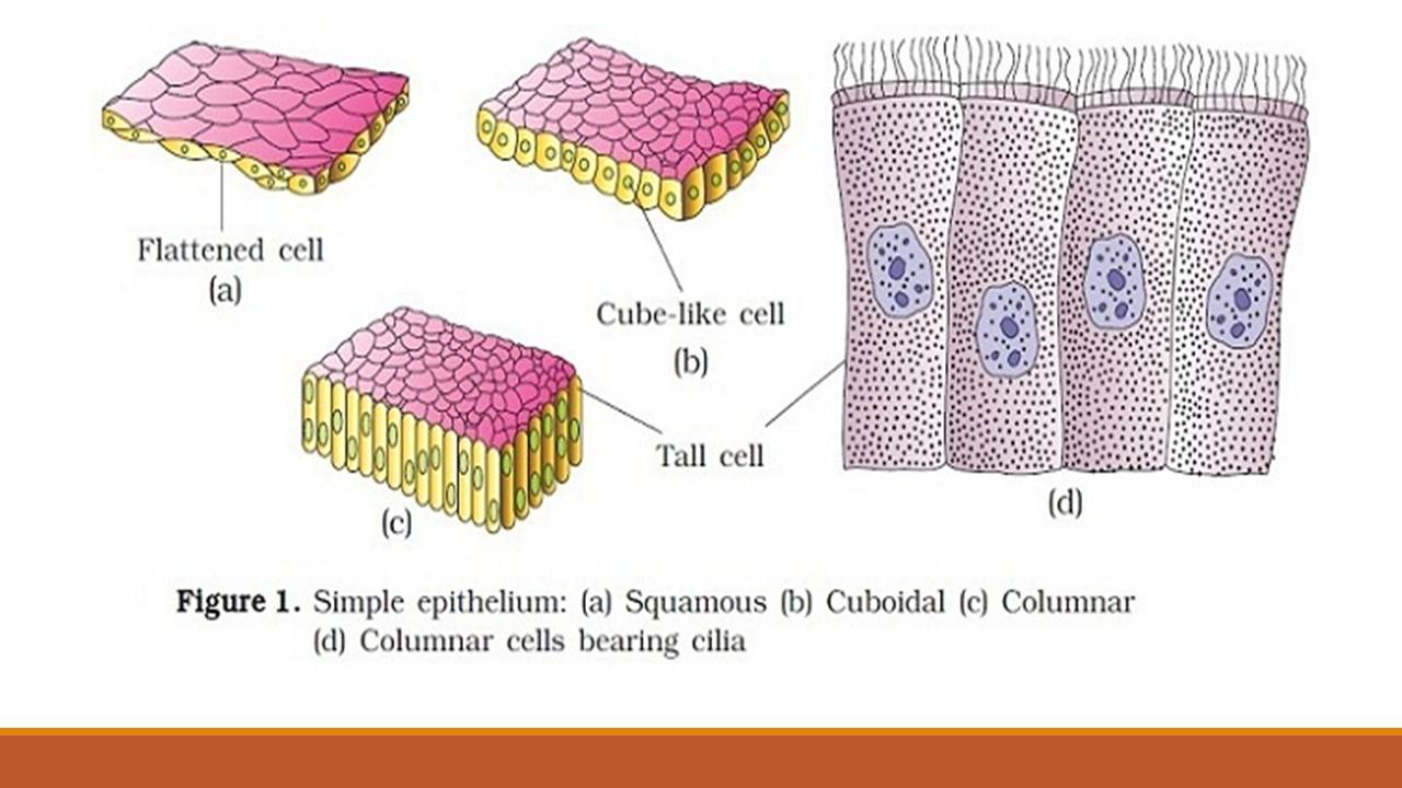
Types of Epithelial Tissue. There are three types of epithelial cells, which differ in their shape and function. Squamous- thin and flat cells Cuboidal- short cylindrical cells, which appear hexagonal in cross-section Columnar- long or column-like cylindrical cells, which have nucleus present at the base On the basis of the number of layers present, epithelial tissue is divided into the simple ...
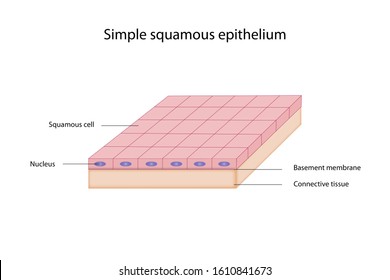
Simple squamous epithelium labeled diagram
Anatomy and Physiology questions and answers. A. Drawings 1. Simple Squamous Epithelium (Example of Epithelium) – label Nucleus, Nucleolus, Plasma Membrane, Cytoplasm 2 Bone (Example of Connective Tissue) - label Central (Haversian) Canal, Concentric Lamellae, Osteocytes in lacunae, Canaliculi, Matrix 4. Giant Multipolar Neuron (Example of ... Skin Diagram Labeling . 1. Label the diagram with the . letters. below according to the structure/area they describe. You may label with a line or put the label directly onto the area described. Be as precise as possible. If you are worried about the precision of your label add a word after to explain exactly where your label should be. What is the structure labeled "5"? (skin diagram) a. arrector pili muscle b. hair matrix c. hair shaft d. inner root sheath e. outer root sheath. d. What is the structure labeled "4"? (skin diagram) ... Blood vessels are lined by a simple squamous epithelium. Skin is made up of a stratified squamous epithelium. In both tissues, the cells are: a ...
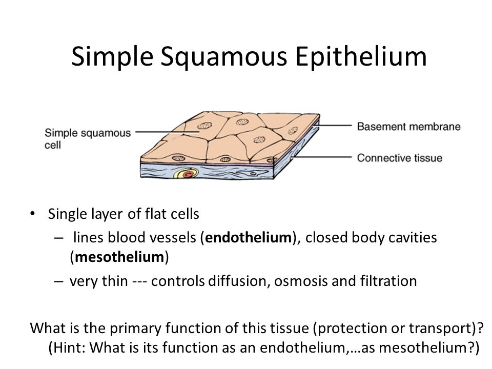
Simple squamous epithelium labeled diagram. The cells found in this epithelium type are flat and thin, making simple squamous epithelium ideal for lining areas where passive diffusion of gases occur. Your goal is to find and learn to recognize simple squamous epithelium on a slide similar to this. The easiest place to find this tissue is the glomerular capsule (don't worry, you don't have to know what that is to find and recognize the tissue). In the top one third of the image you can see ... Simple Squamous Epithelium •Diffusion and Filtration •Secretes lubricating substances in serosae. Identify the structure indicated. Skeletal Muscle Peripherally Located Nucleus. Identify the tissue type and a location where it is found. Skeletal Muscle •Voluntary skeletal muscles attached to bones. Start studying Label the diagram of simple squamous epithelium. Learn vocabulary, terms, and more with flashcards, games, and other study tools.
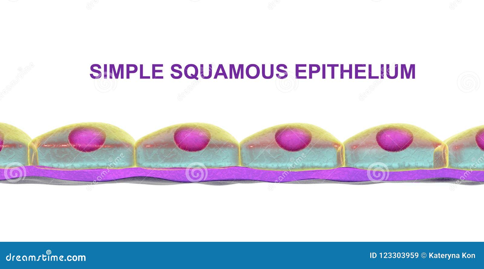
April 29, 2020 - Simple squamous epithelium is a single sheet of cells joined together by flat cells called squamous cells that line the serous cavities of bodies and the Both the endothelial lining of blood vessels and the mesothelial lining of the body cavities are simple squamous epithelium. Try to identify the simple squamous epithelia in these pictures. 2. sep. 2021 ... Figure 1 shows a diagram of simple squamous epithelium labeled. The tissue is polarized with one surface that faces the external environment ... Simple squamous epithelial cells are thin and flat (the thinnest of all epithelial cell-types), which allows them to have a large surface area that is ...
April 9, 2018 - Simple epithelium is one of the types of epithelium that is divided into simple columnar epithelium, simple squamous epithelium, and simple cuboidal epithelium. Bodytomy provides a labeled diagram to help you understand the structure and function of simple columnar epithelium. A basement membrane lies beneath ... and metabolites reach the epithelium by diffusion. Epithelia are polarized, with an apical surface that faces the external environment and a basal surface that faces the basement membrane. ... Simple squamous epithelia consist of a single ... August 12, 1996 - April 29, 2017 - A simple squamous epithelium is a tissue formed from one layer of squamous cells that line surfaces. Squamous cells are large, thin, and flat and contain a rounded nucleus.
Simple squamous epithelium Images, Stock Photos & Vectors ...
Alveoli consist of a layer of simple squamous epithelium resting on a basement membrane. They are intimately associated with a dense network of capillaries. The capillary wall is also made up of simple squamous epithelium resting on a thin basement membrane. Both the layers are thin walled and have similar structure.
Solved] Simple Squamous Epithelium 2. Simple Cuboidal ...
Simple squamous epithelium cells are flat in shape and arranged in a single layer. This single layer is thin enough to form a membrane that compounds can move through via passive diffusion. This epithelial type is found in the walls of capillaries, linings of the pericardium, and the linings ...

Tissue. Tissue classification. Epithelial tissue
Figure 1: Hand-drawn cross section of simple squamous epithelial tissue (not to scale) · Simple squamous epithelial cells function as mediators of filtration and diffusion. Due to their simple and thin construct, they allow for easy transmembrane movement (i.e. across the membrane, and through ...
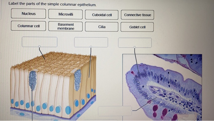
Solved Label the parts of the simple columnar epithelium ...
The structure highlighted with normal color is, in three-dimensions, a sphere composed of a thin outer wall of cells, a space that contains fluid, and an inner region of cells · The outer wall is composed of a single layer of flat cells (a simple squamous epithelium).
Squamous Epithelium High Resolution Stock Photography and ...
Epithelial tissues are classified ... either squamous (flattened and thin), cuboidal (boxy, as wide as it is tall), or columnar (rectangular, taller than it is wide). Similarly, cells in the tissue can be arranged in a single layer, which is called simple epithelium, or more than ...
Simple Squamous Epithelium
text/html; charset=windows-1252; Tutorials on Histology. How to identify tissues (Epithelium, Connective Tissue, Nervous Tissue, Muscle). Includes links to the histology zoomer.

4.2 Epithelial Tissue – Anatomy & Physiology
Simple squamous epithelium definition. Simple squamous epithelium is a type of simple epithelium that is formed by a single layer of cells on a basement membrane. It is a type of epithelium formed by a single layer of squamous or flat cells present on a thin extracellular layer, called the basement membrane.
Epithelium - Wikipedia
Cell shapes can be squamous (flattened and thin), cuboidal (boxy, as wide as it is tall), or columnar (rectangular, taller than it is wide). Similarly, the number of cell layers in the tissue can be one—where every cell rests on the basal lamina—which is a simple epithelium, or more than ...

13 Stratified squamous Epithelium ideas | stratified squamous ...
a) Simple squamous epithelium allows for rapid diffusion. b) The keratin present on simple cuboidal epithelium protects from water loss. c) The microvilli on simple columnar epithelium helps to maximize absorption. d) The keratin on the esophageal stratified squamous epithelium helps to moisten the lumenal surface for swallowing.

BIOL 237 Class Notes - Histology
A simple squamous epithelium, also known as pavement epithelium, and tessellated epithelium is a single layer of flattened, polygonal cells in contact with the basal lamina (one of the two layers of the basement membrane) of the epithelium. This type of epithelium is often permeable and occurs ...
Exercise 4: Epithelium
The Biology Department encompasses areas ranging from molecular biology to ecosystem ecology. This breadth is reflected in our curriculum and in faculty and student research. Our joint mission of teaching and research strongly complement each other and provide students with an educational ...
Simple epithelium: Location, function, structure | Kenhub
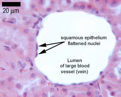
February 6, 2018 - The labels are L for lumen, CT ... simple squamous epithelial cell. Total Magnification: 200x ... Please put the total magnification in the description under the picture, and keep the objective magnification in the title as you have it. Also, when showing a certain type of epithelium, use a bracket ...
How can epithelial tissue be explain perfectly in short way ...
The simple squamous epithelium location specifically exists in the lining of the blood vessels like the arteries, veins, and capillaries. It is also found lining the alveoli or air sacs within the ...
Biology Edited by Dr. Linda Sallah Fawzi
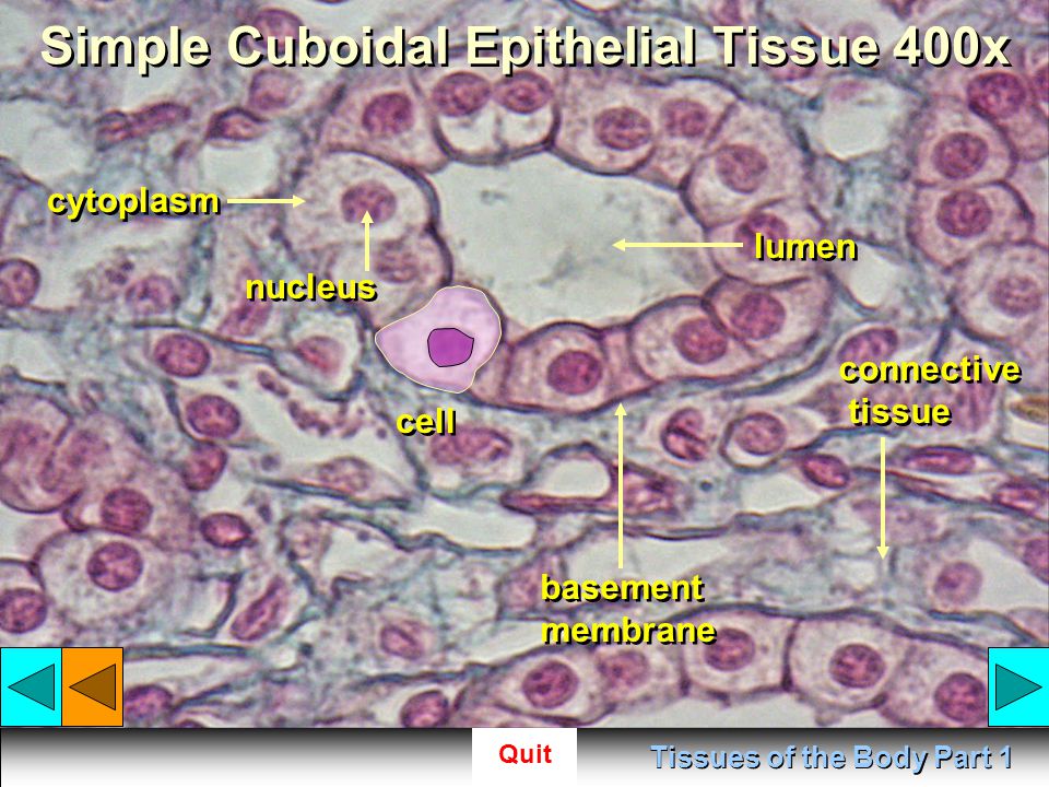
Simple Cuboidal Epithelium. Function: Secretion and Absorption. Location: Glandular tissue, kidney tubules. Simple Columnar Epithelium. Function: Absorption.
Types of epithelial tissue: simple, compound and specialized ...
This is called a stratified squamous epithelium and occurs in the skin and in tissues lining the mouth and vagina. ... Cuboidal epithelial cells, shown in Figure 2, are cube-shaped with a single, central nucleus. They are most commonly found in a single layer representing a simple epithelia ...

Lab 2 Epithelial tissue | Histology
What is the structure labeled "5"? (skin diagram) a. arrector pili muscle b. hair matrix c. hair shaft d. inner root sheath e. outer root sheath. d. What is the structure labeled "4"? (skin diagram) ... Blood vessels are lined by a simple squamous epithelium. Skin is made up of a stratified squamous epithelium. In both tissues, the cells are: a ...

Simple squamous epithelium - Wikipedia
Skin Diagram Labeling . 1. Label the diagram with the . letters. below according to the structure/area they describe. You may label with a line or put the label directly onto the area described. Be as precise as possible. If you are worried about the precision of your label add a word after to explain exactly where your label should be.
Types of epithelial tissue: simple, compound and specialized ...
Anatomy and Physiology questions and answers. A. Drawings 1. Simple Squamous Epithelium (Example of Epithelium) – label Nucleus, Nucleolus, Plasma Membrane, Cytoplasm 2 Bone (Example of Connective Tissue) - label Central (Haversian) Canal, Concentric Lamellae, Osteocytes in lacunae, Canaliculi, Matrix 4. Giant Multipolar Neuron (Example of ...
Simple Squamous Epithelium - Definition and Examples ...
Epithelium Web Lab
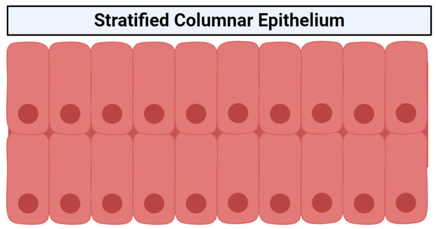
Stratified columnar epithelium- structure, functions, examples

Simple Columnar Epithelium
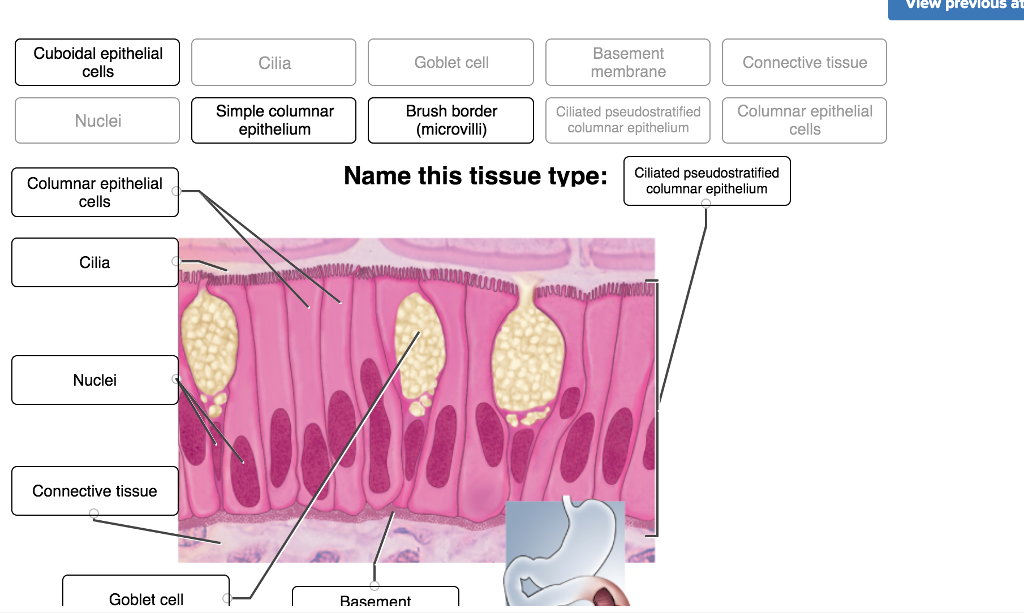
Solved Vlew previous atten Simple Simple squamous | Chegg.com

Stratified squamous epithelium - Wikipedia
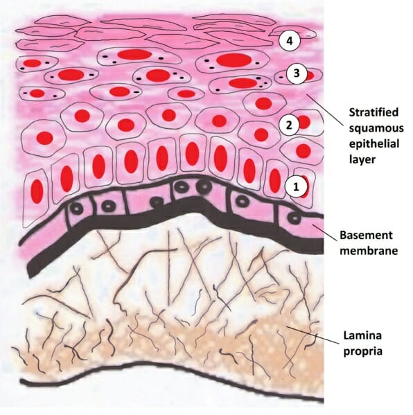
Stratified Squamous Epithelium - Definition and Function ...
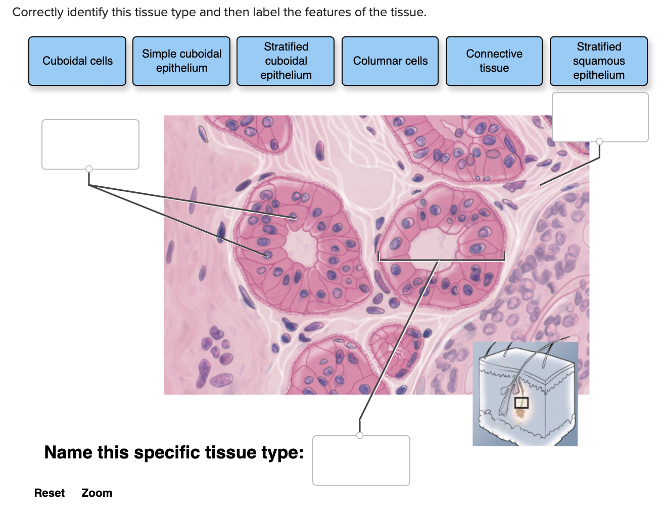
Solved Correctly identify this tissue type and then label ...
Types of epithelial tissue: simple, compound and specialized ...
Simple Squamous Epithelium Stock Illustrations – 11 Simple ...
Epithelia: The Histology Guide
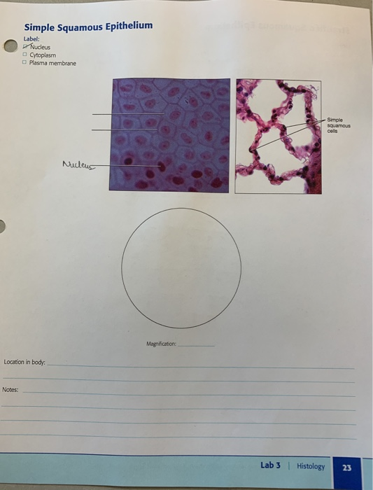
Solved Simple Squamous Epithelium Label: Nucleus Cytoplasm ...

Epithelia: The Histology Guide

Examining epithelial tissue under the microscope | Human ...

48 Tissues ideas | tissue, anatomy and physiology, histology ...

290 Squamous Epithelium Photos and Premium High Res Pictures ...

Stratified Squamous Epithelium Diagram | Quizlet

Simple squamous epithelium - Definition and Examples ...

How to draw simple squamous epithelium @Bigyan - The Science || most easy way

4.2 Epithelial Tissue – Anatomy & Physiology

Epithelium Lab

Simple Cuboidal Epithelium: Location, Structure & Function ...
0 Response to "40 simple squamous epithelium labeled diagram"
Post a Comment