41 wiggers diagram aortic regurgitation
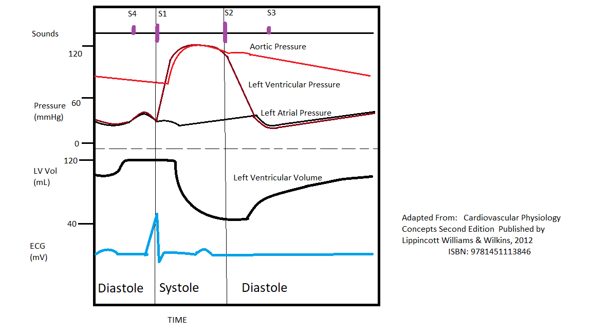
Why you might avoid beta blockade in severe aortic ... Read more: Aortic Regurgitation in Chapter 283: Aortic Valve Disease, Harrison's Principles of Internal Medicine 19e. The above Wiggers diagrams are modifications of: adh30 revised work by DanielChangMD who revised original work of DestinyQx; Redrawn as SVG by xavax - Wikimedia Commons: Wiggers Diagram.svg, CC BY-SA 4.0 « What causes alcohol withdrawal seizures? [5+] Download Wiggers Diagram And The Description ... Blood entering the ventricles mitral stenosis aortic regurgitation. Wiggers diagrams can vary in detail and number of variables presented. The Wiggers diagram is a synchronous tracing of aortic pressure left atrial pressure left ventricular pressure left ventricular volume and EKG throughout the cardiac cycle.
The Cardiac Cycle - What is Mitral Valve Regurgitation Aortic/Pulmonary artery pressure increases slightly during this phase due to blood pushin back against the now closed aortic/pulmonary valve. Figure 2: (a) The Wigger's diagram, which shows changes...

Wiggers diagram aortic regurgitation
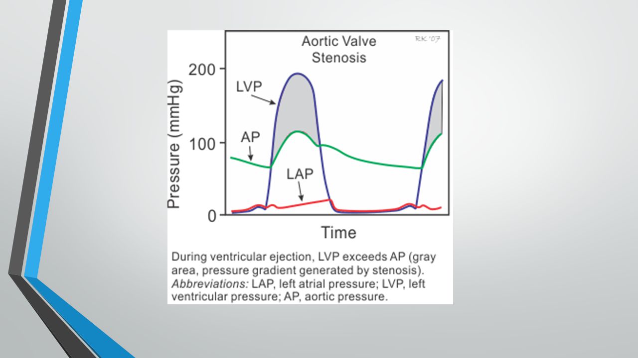
Wiggers Diagram Aortic Stenosis - wiringall.com If a high-pressure jet resulting from aortic stenosis is present, the aorta will stretch If we go back to Part I and look at the Wigger's diagram, the aortic valve is. Senile calcific aortic stenosis: age range and gender difference . Changes in the pressure-volume loop and murmur on a wiggers diagram: Mitral stenosis. 38 wiggers diagram quiz - fmcggyartok.blogspot.com The diagram uses the left chambers of the heart to demonstrate: Aortic pressure Atrial pressure Ventricular pressure Ventricular volume Electrocardiogram (ECG) Phonocardiogram (heart sounds) Wiggers Diagram Mar 05, 2002 · The first set of questions deals with the the events during each of the phases or periods of the normal cardiac cycle. Valvular Stenosis - CV Physiology Aortic valve stenosis is characterized by the left ventricular pressure being much greater than aortic pressure during left ventricular (LV) ejection (see figure at right). In this example, LV peak systolic pressure during ejection is 200 mmHg (normally ~120 mmHg) and the aortic pressure is slightly reduced to from 120 to 110 mmHg.
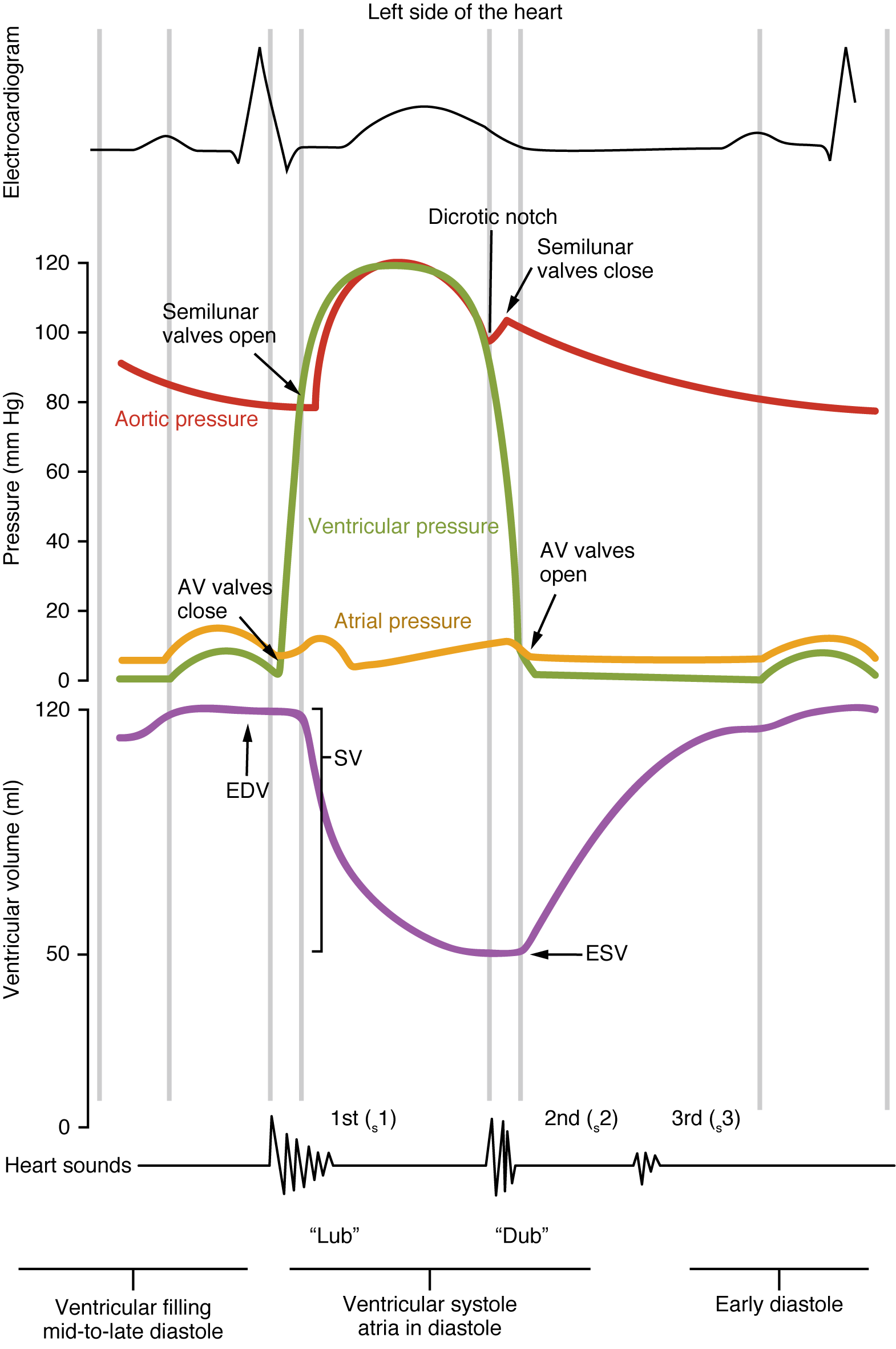
Wiggers diagram aortic regurgitation. The Wright table of the cardiac cycle: a ... - Physiology A typical Wiggers diagram is shown in Fig. 1. Fig. 1. The Wiggers diagram. From top to bottom, the lines show: 1) aortic pressure, 2) ventricular pressure, 3) atrial pressure, 4) electrocardiogram, 5) mitral and aortic valve opening and closing, and 6) heart sounds. The y -axes vary, but all share a common x -axis in time. 4: Arterial pressure | Thoracic Key Figure 4.1 Schematic of aortic pressure. Figure 4.2 Wiggers diagram [LVEDV = Left Ventricular End Diastolic Volume; LVESV = Left Ventricular End Systolic Volume]. Figure 4.3 Aortic pressure in a hypovolemic patient. Aortic pressure tracings taken in a patient before and after an intravenous (IV) fluid bolus. Free Nursing Flashcards about Wiggers Diagram In early ventricular systole. The semilunar valves open. Ventricular Diastole represents. The period of time the ventricles are filling. Between the end of the T wave and the onset of the QRS complex. Ventricular Diastole. Approximately 70% of the Diastolic filling occurs during. Early Ventricular Diastole. Doppler Echocardiography Unraveling Lewis and Wiggers Diagrams Diastolic mitral regurgitation (DMR) has been reported in patients with AV block, aortic regurgitation, cardiomyopathies, and in patients with long filling periods in atrial tachyarrhythmias.
8-28 Wigger plot review + mitral valve prolapse ... - Quizlet What elements of the wigger diagram will look different in mitral regurgitation large v wave and rapid y descent A patient is complaining of trouble with breathing - you auscultate and hear a murmur occurring throughout systole, turbulent flow during diastole, and S3. Wiggers Diagram Aortic Regurgitation - schematron.org Diagram of the ascending and descending aorta illustrating . Stewart10 and later Wiggers and Green Aortic insufficiency (AI), also known as aortic regurgitation (AR), is the leaking of the aortic valve of the heart that causes blood to flow in the reverse direction. Wiggers Diagram Aortic Stenosis - schematron.org If a high-pressure jet resulting from aortic stenosis is present, the aorta will stretch If we go back to Part I and look at the Wigger's diagram, the aortic valve is. Dr. Carl Wiggers' careful observations have provided a meaningful resource for students to learn how the heart works. Throughout the many years from his initial .Valvular Stenosis. Valvular Insufficiency (Regurgitation) - CV Physiology Aortic regurgitation occurs when the aortic valve fails to close completely and blood flows back from the aorta (Ao) into the left ventricle after ejection into the aorta is complete and during the time that the left ventricle (LV) is also being filled from the left atrium (LA) (see figure at right). Because the ventricle is being filled from two sources (aorta and LA), this leads to much ...
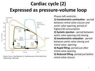
PDF Wiggers, Diagrams and Heart Sounds - University of Michigan Wigger's diagram: This seemingly complex diagram (Don't Panic!) is actually a very useful tool to learn the components of the cardiac cycle and how they relate to cardiac exam findings, which I have indicated in blue. To start, identify the aortic, LV and LA pressure lines and talk yourself through the sequence of Do You Have Any Bicuspid Aortic Valve Pictures? - Adam's ... Well, considering I had a bicuspid aortic valve for the first 33 years of my life, I definitely researched and located pictures of bicuspid aortic valves for my book. As you can see below, the aortic valve on the left is normal - the valve has three leaflets and three cusps. The valve on the right, however, is a bicuspid aortic valve. As you ... PDF Pressure-Volume Loop: Ventricular Physiology and Pathology 27. Can we use the term "afterload" for aortic pressure during the phase that the aortic valve is closed? _____ _____ 28. Above diagram shows the relationship of Frank-Starling curves to afterload and stroke volume. The centrally located curve marked "B" is for normal heart. Which of the two curves; A or C, represents increased and ... Wigger's Diagrams: Aortic Stenosis, Aortic Insufficiency ... About Press Copyright Contact us Creators Advertise Developers Terms Privacy Policy & Safety How YouTube works Test new features Press Copyright Contact us Creators ...
Chronic kidney disease and valvular heart disease ...
Wiggers Diagram Aortic Regurgitation - Wiring Diagrams Wiggers Diagram Aortic Regurgitation findings of aortic insufficiency consisting of a wide .. Diagram of the ascending and descending aorta illustrating . Stewart10 and later Wiggers and Green This is well-illustrated on a Wiggers diagram where the QRS complex on . blood entering the ventricles (mitral stenosis, aortic regurgitation).

Pressure volume diagram - wikidoc
wiggers diagram and diastolic pressure : Cardiology wiggers diagram and diastolic pressure. Hi, I know that systolic pressure is the highest pressure in the left ventricle during systole, but why does ventricular pressure (bright blue line) fall to near zero in the diastolic part of wiggers diagram? I thought DBP should hover around 80 not zero? sorry if this is not the right forum for posting ...

The Cardiac Cycle - What is Mitral Valve Regurgitation
Physiology, Cardiac Cycle - StatPearls - NCBI Bookshelf This rhythmic sequence causes changes in pressure and volume that are often seen graphically in the form of a Wiggers diagram or venous pressure tracings. Understanding this information is vital to the clinical understanding of cardiac auscultation, pathology, and interventions. Cellular
Physiology, Cardiac Cycle Article
PDF The Cardiac Cycle - University of Cape Town The "Wiggers diagram" ... Mitral and aortic regurgitation are the most common regurgitant lesions. Alternatively, the orifice of a valve may become narrowed or stenotic. This obstructs the flow of blood through it and requires increased pressure gradients to be generated across the valve to achieve

Cardiac physiology - Knowledge @ AMBOSS
PDF The Cardiac Cycle Understand the Wiggers Diagram of Cardiac Cycle Differentiate between Wiggers Diagram and the Pressure Volume Curve ... Right Ventricle Pulmonary Artery Left Atria Left Ventricle Aorta. Valves: Atrioventricular Tricuspid Valve Mitral Valve Semilunar Pulmonary Valve Aortic Valve. 130/80 2 8 30/6 130/10 25/8 Pressures: Right Atria (2) Right ...

Split S2 - Wikipedia
Wiggers Diagram Tutorial - XpCourse Wiggers diagram A Wiggers diagram is a standard diagram used in cardiac physiology named after Dr. Carl J. Wiggers. The X axis is used to plot time, while the Y axis contains all of the following on a single grid: More Courses ›› View Course (PPT) Ciclo cardiaco y diagrama de wiggers | Gabriela ... Now

Wigger's Diagram Quiz
Heart Murmurs | Clinical Features - Geeky Medics Aortic regurgitation. Aortic regurgitation (AR) occurs when there is backflow of blood from the aorta into the left ventricle during ventricular diastole. Aortic regurgitation is associated with an early diastolic murmur heard loudest at the left sternal edge Aetiology. Aortic regurgitation can be either acute or chronic. Chronic AR is often ...
Normal Circulation and Congestive Heart Failure
PV Loops, Wigger's Diagram & Starling Curve Flashcards ... Aortic Regurgitation Wigger's Diagram ↓↓Aortic pressure during diastoleole* -blood leaking out of aorta faster than usual -Wider pulse pressure Venous Pressure Tracings -represent pressure changes in jugular vein, SVC, RA, pulmonary vein & LA 1. A Wave=RAcontraction (↑pressure, RV filling) 2.
Adult Echocardiography Review - ppt download
PDF IN FURTHER OBSERVATIONS ON EXPERIMENTAL AORTIC INSUFFICIENCY II. CINEMATOGRAPHIC STUDIES OF CHANGES IN VENTRICULAR SIZE AND IN LEFT VENTRICULAR DISCHARGE By CARLJ. WIGGERS, HAROLD THEISENAND HARLEYA. WILLIAMS (FromtheDepartmentofPhysiology, WesternReserve University MedicalSchool, Clevdand) (Receivedforpublication April30, 1930) INTRODUCTION

Wigger's Diagrams: Aortic Stenosis, Aortic Insufficiency, Mitral Stenosis, Mitral insufficiency
Physiology, Cardiac Cycle Article - StatPearls This rhythmic sequence causes changes in pressure and volume that are often seen graphically in the form of a Wiggers diagram or venous pressure tracings. Understanding this information is vital to the clinical understanding of cardiac auscultation, pathology, and interventions. Cellular

Valve cases Flashcards | Quizlet
Valvular Stenosis - CV Physiology Aortic valve stenosis is characterized by the left ventricular pressure being much greater than aortic pressure during left ventricular (LV) ejection (see figure at right). In this example, LV peak systolic pressure during ejection is 200 mmHg (normally ~120 mmHg) and the aortic pressure is slightly reduced to from 120 to 110 mmHg.

loadbinarycawul2k2.gif (684×895) | Cardiology, Vet med, Medicine
38 wiggers diagram quiz - fmcggyartok.blogspot.com The diagram uses the left chambers of the heart to demonstrate: Aortic pressure Atrial pressure Ventricular pressure Ventricular volume Electrocardiogram (ECG) Phonocardiogram (heart sounds) Wiggers Diagram Mar 05, 2002 · The first set of questions deals with the the events during each of the phases or periods of the normal cardiac cycle.

Lecture 21 Adult Echocardiography Final Exam Prep 2 - ppt ...
Wiggers Diagram Aortic Stenosis - wiringall.com If a high-pressure jet resulting from aortic stenosis is present, the aorta will stretch If we go back to Part I and look at the Wigger's diagram, the aortic valve is. Senile calcific aortic stenosis: age range and gender difference . Changes in the pressure-volume loop and murmur on a wiggers diagram: Mitral stenosis.
UNIVERSITÀ DEGLI STUDI DI PAVIA FACOLTÀ DI INGEGNERIA CORSO ...

Hemodynamics in the Cardiac Catheterization Laboratory of the ...
Why is there back flow of blood (dicrotic notch) when aortic ...

CV Physiology | Valvular Insufficiency (Regurgitation)
Controlling the Kinematics of the Left Ventricle to Treat ...
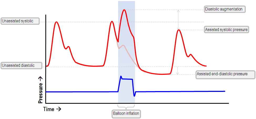
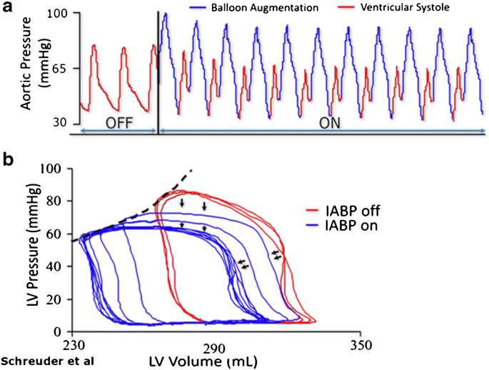
The normal IABP waveform | Deranged Physiology

CV Week 3 Flashcards | Quizlet
Aortic Regurgitation | Washington Manual of Medical Therapeutics
5: Wiggers diagram for the left heart. | Download Scientific ...

Chronic kidney disease and valvular heart disease ...

Pin on KAPLAN QBANK HEART

pump quiz
Cardiac cycle and heart sounds | Cardiovascular system ...

Wigger's Diagram | Draw it to Know it
Cardiac Cycle
Physiologic and pathophysiologic function of the heart

CGI animated graphic of the human heart, sectioned, with ...

The Wright table of the cardiac cycle: a stand-alone ...

29 RCIS - waveforms ideas | cardiac nursing, icu nursing ...

Why you might avoid beta blockade in severe aortic ...

Wiggers diagram. The diagram shows the aortic, atrial and ...

Wiggers diagram - Wikipedia
Modified Wiggers diagram. A sample of the SCG signal in the ...

Aortic regurgitation - Knowledge @ AMBOSS

Tumblr
Mechanical Support in Early Cardiogenic Shock: What Is the ...
Pressure–volume loop analysis in cardiology - Wikiwand

Hemodynamics in the Cardiac Catheterization Laboratory of the ...
0 Response to "41 wiggers diagram aortic regurgitation"
Post a Comment