41 hip flexor anatomy diagram
What Are the 3 Main Hip Flexor Muscles? | Livestrong.com The hip flexor muscles are attached to the hip joint to allow the femur, which is the upper leg bone, to flex onto the pelvis region. In simpler terms, the hip flexor muscles allow the knee to raise and move the thigh upward. The hip is a large, deep and stable ball and socket joint that is surrounded by many ligaments, tendons and muscles. Understanding Hip Flexor Pains | What Causes Pain in Hip ... Avascular necrosis. This is a very rare hip flexor pain. It is caused when blood supply to a bone is completely cut off or significantly reduced. This type of hip pain is often felt in the center of the groin, thigh, or buttocks. It can develop gradually or can be caused by trauma, or excessive alcohol or steroid use.
Unlock Your Hip Flexors: HumanampAnimal Anatomy and ... leg (anatomy) Leg, limb or appendage of an animal, used to support the body, provide locomotion, and, in modified form, assist in capturing and eating prey (as in spiders and insects). In four-limbed vertebrates all four appendages are commonly called legs, but in bipedal animals only the lower two are so called. H b ️Nerve Chart

Hip flexor anatomy diagram
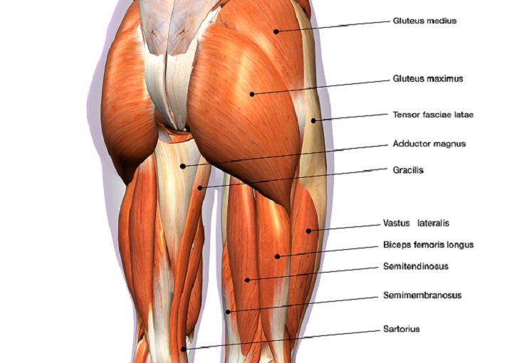
› en › libraryHip and thigh muscles: Anatomy and functions | Kenhub Small and deep muscles which mainly externally rotate the thigh at the hip joint and stabilize the pelvis. These are the piriformis, obturator internus, obturator externus, gemellus superior, gemellus inferior, and quadratus femoris. They are also known as the inner hip muscles and deep external rotators. Superficial layer Hip Flexor Muscles | Anatomy Of The 5 Major Hip Flexors ... A hip flexor muscle is a muscle that functions in flexing the hip - in other words in bringing the knee closer to the chest. This post discusses the 5 major hip flexor muscles and their anatomy - Psoas, Iliacus, Rectus Femoris, Tensor Fasciae Latae, and Sartorius.. Hip Flexor Muscle #1: Psoas. The Psoas muscle is a powerful, deep hip flexor that connects from the lumbar vertebrae to the top of ... Gallery of hip flexor muscles anatomy anatomy diagram book Hip Flexor Muscles Anatomy Anatomy Diagram Book images that posted in this website was uploaded by Feeds.canoncitydailyrecord.com. Hip Flexor Muscles Anatomy Anatomy Diagram Book equipped with a HD resolution x .You can save Hip Flexor Muscles Anatomy Anatomy Diagram Book for free to your devices.. If you want to Save Hip Flexor Muscles Anatomy Anatomy Diagram Book with original size you can ...
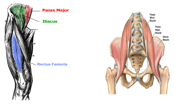
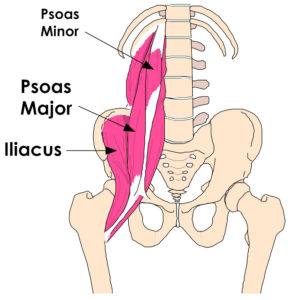
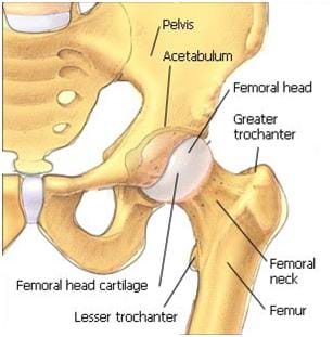
Hip flexor anatomy diagram. Basic Anatomy of Stretching the Hip Flexors - Movement Fix This week we are returning to our discussion of 'the basic anatomy' series and we are taking a look at the hip flexors, but specifically the psoas and iliacus muscles. The hip flexors are all the muscles that flex the hip joint. Surprise! There are 4: sartorius, rectus femoris, iliacus, and psoas. Hip joint: Bones, movements, muscles - Kenhub Hip joint (Articulatio coxae) The hip joint is a ball and socket type of synovial joint that connects the pelvic girdle to the lower limb. In this joint, the head of the femur articulates with the acetabulum of the pelvic (hip) bone.. The hip joint is a multiaxial joint and permits a wide range of motion; flexion, extension, abduction, adduction, external rotation, internal rotation and ... Overview of Hip Flexor Muscles and Injuries The hip flexors are several muscles that bring your legs and trunk together in a flexion movement. They allow you to move your leg or knee up towards your torso, as well as to bend your torso forward at the hip. You can strain or tear your hip flexor muscles through sudden movements or falls. 1 Jan-Otto / E+ / Getty Images Anatomy and Function Hip Anatomy Diagram: From Bones To Joints - Science Trends General Hip Anatomy. The hip is a ball-and-socket joint, similar to the joint in the shoulder. Part of the reason for the hip's stability is that there is a very deep socket, called the acetabulum, in the hip joint. A strong capsule joint supported by ligaments and muscles also provides extra stability to the hip.
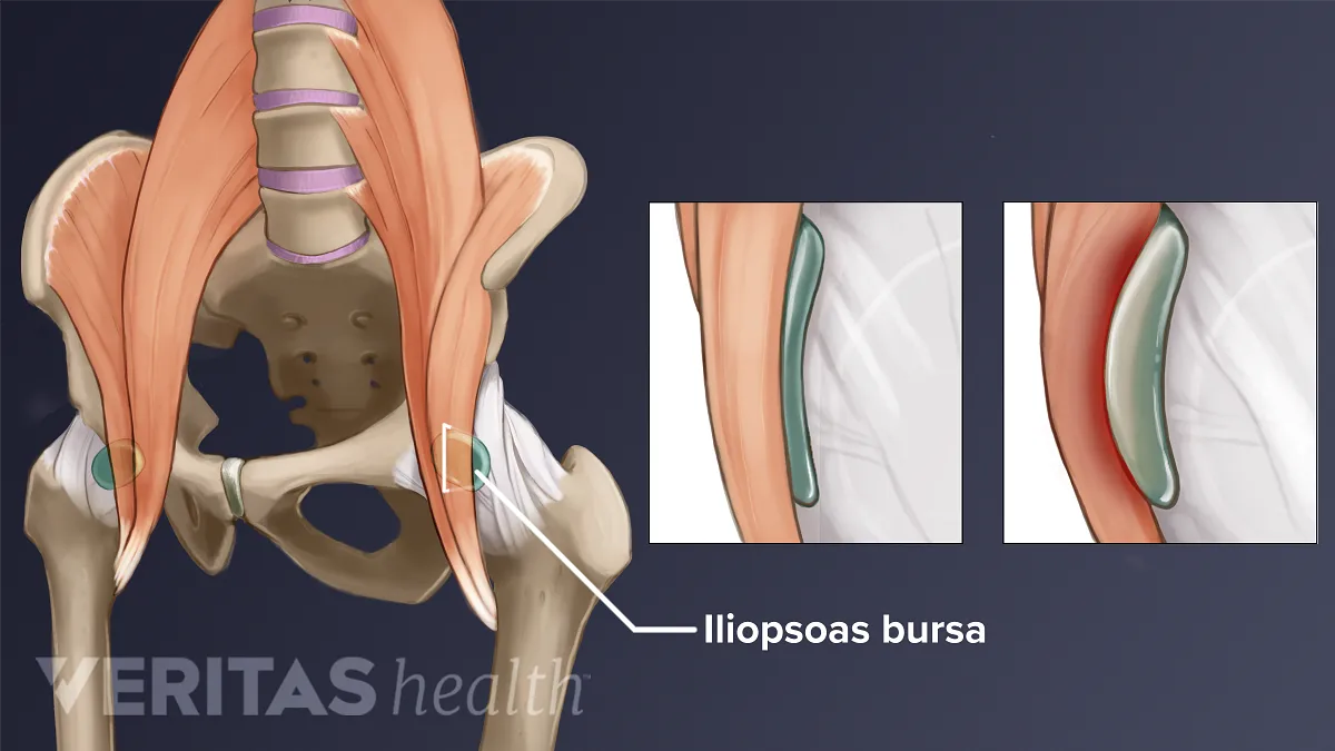
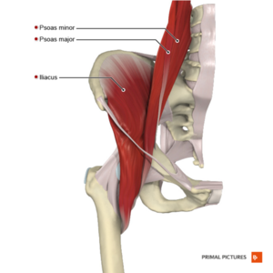
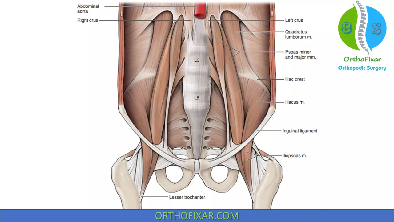
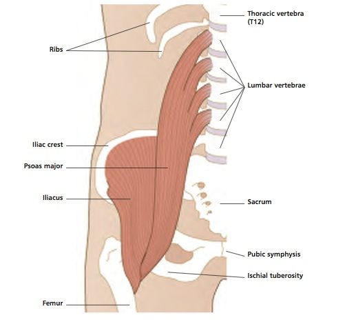
Iliopsoas Muscle: Anatomy, Function, and Treatment The iliopsoas muscle is a major mover of your hip joint. It's formed by the joining of three muscles: the iliacus muscle, the psoas major muscle, and the psoas minor muscle. These muscles work together to flex your hip and to stabilize your hip and lower back during activities such as walking, running, and rising from a chair. A Guide to Hip Anatomy: Bones, Muscles, Tendons & Pain ... Adductor muscles on the inside of your thigh. Iliopsoas muscle, a hip flexor muscle that attaches to the upper thigh bone. Rectus femoris muscle, one of the quadriceps muscles on the front of your... Hip Anatomy, Pictures, Function, Problems & Treatment The hip joint is a ball-and-socket type joint and is formed where the thigh bone (femur) meets the pelvis. The femur has a ball-shaped head on its end that fits into a socket formed in the pelvis, called the acetabulum. Large ligaments, tendons, and muscles around the hip joint hold the bones (ball and socket) in place and keep it from dislocating. Hip Flexor Stretches to Counter the Effects of Sitting ... If done with proper alignment, Warrior Pose I can be a wonderful hip flexor stretch. Stand with one leg forward and one leg back, ready for Warrior I. Put your fingers on the front pelvis bones: You should be able to feel a small, round protuberance on each side, called the anterior superior iliac spine, or ASIS.
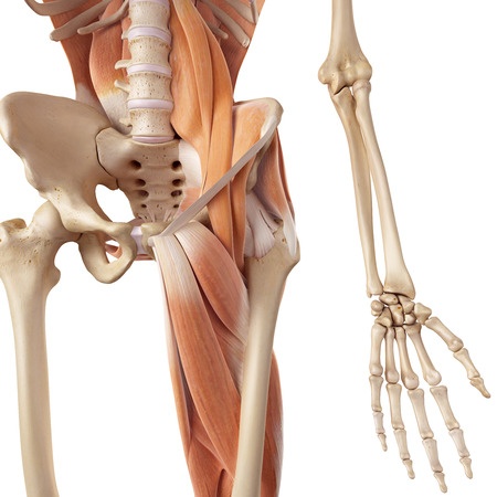
Hip Flexor Muscles and Anatomy for Personal Trainers All of the hip flexor muscles attach from the pelvis or spine to the femur or tibia, which is how they influence hip flexion. Bypass the tricky bony landmark terms for now and familiarize yourself with just the two bones each muscle attaches to. The bolded words in the descriptions below are there just for you intermediate anatomy student! TFL, IT Band & Hip Flexors Diagram | Quizlet Start studying TFL, IT Band & Hip Flexors. Learn vocabulary, terms, and more with flashcards, games, and other study tools. hippainhelp.com › anterior-hip-painAnterior Hip Pain - Pain at the front of the hip Sep 06, 2019 · Anterior hip pain is pain that is experienced over the front of the hip. There are many potential causes for anterior hip pain. Explore the information under each tab below to understand more about the anatomy of the area and things that may go wrong. Anatomy of the Hip Flexor Muscles - Iliacus and the Psoas ... Anatomy of the Hip Flexor Muscles - Iliacus and the Psoas Major, Iliopsoas, Rectus Femoris Hip Flexor Muscle Anatomy The Iliopsoas actually consists of two muscles: the Iliacus and the Psoas Major. Together, they are known as the Iliopsoas. Anatomy Chart courtesy of FCIT The Iliacus originates on the pelvic crest and attaches on the femur.
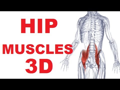
Deep Dive into the Anatomy of the Hip Flexor Muscles
Where Is Your Hip Flexor? - Body Pain Tips The hip flexor has both major and minor functions, it is able to fulfill different roles because it is composed of several muscles, the largest ones are discussed below. The primary goal of the hip flexor is to facilitate flexion of the hip joint. In normal terms, this means that the hip flexor is used anytime the knee is lifted up, a step is ...

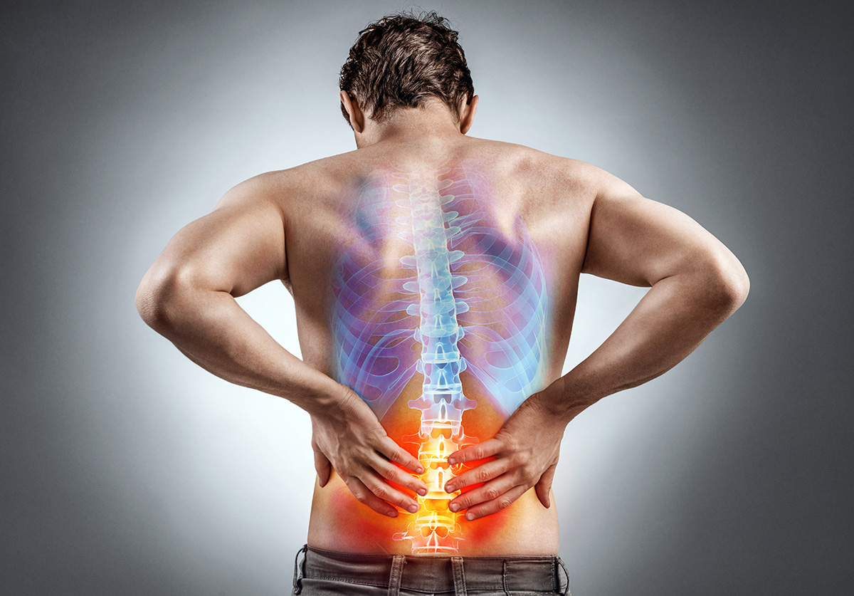
Pin on Back Pain
Anatomy hip muscles Images, Stock Photos & Vectors ... Anatomy hip muscles images. 6,324 anatomy hip muscles stock photos, vectors, and illustrations are available royalty-free. See anatomy hip muscles stock video clips. Image type.

Psoas Hip-Flexors - Home | Facebook
Hip flexor strain: Symptoms, recovery time, treatment, and ... The hip flexors connect the top of the femur, which is the largest bone in the body, to the lower back, hips, and groin. There are various hip flexor muscles that all work to enable a person to...
Hip flexors Images, Stock Photos & Vectors | Shutterstock
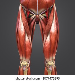
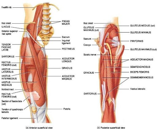
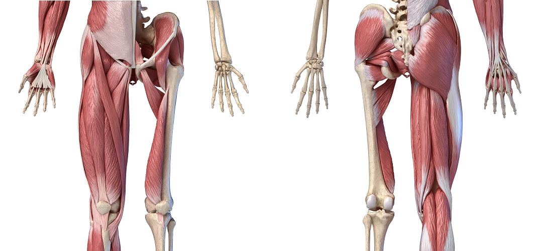
Muscles of the Hip - Anatomy Pictures and Information These muscles can be grouped based upon their location and function. The four groups are the anterior group, the posterior group, adductor group, and finally the abductor group. The anterior muscle group features muscles that flex (bend) the thigh at the hip. Continue Scrolling To Read More Below... Click To View Large Image Continued From Above...

Hip Flexors & Lateral Tight Diagram | Quizlet
PDF Applied anatomy of the hip and buttock rotation at the hip and 90° flexion at the knee, the muscle becomes prominent and is easily palpable. The rectus femoris combines movements of flexion at the hip and extension at the knee. Its origin is at the anterior . inferior iliac spine, a groove above the acetabulum and the fibrous capsule of the hip joint and inserts into the common

Hip Muscles - The Definitive Guide | Biology Dictionary
› en › libraryMuscles of the trunk: Anatomy, diagram, pictures | Kenhub Feb 22, 2022 · Ventral trunk muscles (overview) The trunk (torso) is the central part of the body to which the head and the limbs are attached. Except for the brain, the trunk houses all the vital organs of the human body.

Hip Flexor Ability Anatomy Review [Psoas, Rectus Femoris ...
Diagram Of Hip Anatomy, General Hip Anatomy They include the brain, heart, lungs, spleen, muscles, stomach, kidneys and more. Sartorius is a unique muscle because it is the only knee flexor that originates anteriorly. Bones diagram of the hip bone diagrams of hip bone diagram of hip bones images anatomy structure. Picture of the female body 744 992 diagram - picture of the female body ...
Hip Muscles - The Definitive Guide | Biology Dictionary
What Is a Hip Flexor Strain and How Is It Treated? - NY ... Most hip strain injuries begin as a microscopic tear that gradually increases in size with repetitive hip movement. Addressing a minor tear early on is the best way to prevent the condition from worsening. Symptoms of Hip Flexor Strain. The primary symptom of a hip flexor strain is pain where your thigh meets your hip.

Hip flexor pain. #tiktokpartner #learnontiktok #sportsinjury #pittsburgh #chiropractor
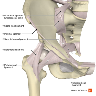
Hip Pain Explained - including structures & anatomy of the ... the joints of the hip & pelvis - hip joint, sacro-iliac joints, pubic symphysis the soft tissues - muscles, tendons, bursae & fascia bones - the femur (thigh bone) or bones of the pelvis local nerves running through and around the hip & pelvis musculoskeletal problems elsewhere in the body, such as the lower back (referred pain)

How Can Tight Hip Flexors Give You a Headache? | Tight hip ...
Muscle Anatomy Diagram Diagram | Quizlet Always doing the opposite. Muscles are located on opposite sides of the bone it is attached to. Examples are: Biceps-Triceps, Hamstring-Quadraceps, Right Obliques-Left Obliques, Abdominals-Erector Spinae, Gluteus Maximus- Hip Flexors, Adductors-Abbductors, Tibialis Anterior-Gastrocnemius, Trapezius-Pectorals, Latissimus Dorsi-Deltoids

Hip Flexor Pain
Hip Flexor Strain: Causes, Symptoms, and Treatment Hip flexor strain occurs when you use your hip flexor muscles and tendons too much. As a result, the muscles and tendons become inflamed, sore, and painful. Some people are more likely than others ...
Causes of Hip Flexor Pain
Gallery of hip flexor muscles anatomy anatomy diagram book Hip Flexor Muscles Anatomy Anatomy Diagram Book images that posted in this website was uploaded by Feeds.canoncitydailyrecord.com. Hip Flexor Muscles Anatomy Anatomy Diagram Book equipped with a HD resolution x .You can save Hip Flexor Muscles Anatomy Anatomy Diagram Book for free to your devices.. If you want to Save Hip Flexor Muscles Anatomy Anatomy Diagram Book with original size you can ...
Hip Flexor Muscles and Anatomy for Personal Trainers
Hip Flexor Muscles | Anatomy Of The 5 Major Hip Flexors ... A hip flexor muscle is a muscle that functions in flexing the hip - in other words in bringing the knee closer to the chest. This post discusses the 5 major hip flexor muscles and their anatomy - Psoas, Iliacus, Rectus Femoris, Tensor Fasciae Latae, and Sartorius.. Hip Flexor Muscle #1: Psoas. The Psoas muscle is a powerful, deep hip flexor that connects from the lumbar vertebrae to the top of ...

Deep Dive into the Anatomy of the Hip Flexor Muscles
› en › libraryHip and thigh muscles: Anatomy and functions | Kenhub Small and deep muscles which mainly externally rotate the thigh at the hip joint and stabilize the pelvis. These are the piriformis, obturator internus, obturator externus, gemellus superior, gemellus inferior, and quadratus femoris. They are also known as the inner hip muscles and deep external rotators. Superficial layer
Hip Flexors - Physiopedia
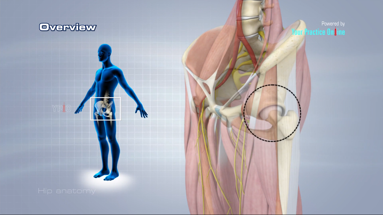
Hip Anatomy Video | Hip Orthopaedics Videos | Your Practice ...
Hip Anatomy - Physiopedia
How do you know you have a hip flexor strain?
Hip and thigh muscles: Anatomy and functions | Kenhub
Hip Muscles - The Definitive Guide | Biology Dictionary
5 Hip Flexor Stretches!

Just Muscles Functional Hip Flexor Muscles
Hip Flexor Tightness and Why It Is Killing Your Low Back ...

5 Minute Reads: Understand the Hip Flexors

Six rotation movements on the X, Y, and Z axes of the hip ...

How To Unlock Tight Hip Flexors - EMPOWER YOUR WELLNESS

Hip Pain Explained - including structures & anatomy of the ...
Hip Anatomy - Iliopsoas Muscle
Hip Muscles Anatomy • Easy Explained - OrthoFixar 2022

HIP FLEXOR STRETCH
Tight Hip Flexors and Back Pain, Knee and Foot Pain » The ...

Hip Flexors - an overview | Hip flexor, Femoral nerve ...
Vivian Grisogono - ABOUT THE HIP

Psoas major Part I: hip flexor or lumbar stabilizer?
Why does the front of my hip pinch?

Movements in anatomical planes From Fig. 4, the movements of ...

hip flexors – Sam Sherrington – Osteopath
Hip and thigh muscles: Anatomy and functions | Kenhub

Leg Anatomy | All About the Leg Muscles
Hip Strains - OrthoInfo - AAOS
Muscles of the hip - Wikipedia
0 Response to "41 hip flexor anatomy diagram"
Post a Comment